Traumatic vertebrobasilar pseudoaneurysms: diagnostic pitfalls on CT angiography with forensic implications — two case reports
The first case illustrated a fundamental limitation of conventional CTA. A PICA pseudoaneurysm was obscured on 5-mm acquisitions but identified retrospectively on 0.5-mm reconstructions with VRT. This finding supports prior study reporting pooled sensitivity was 64% (53–74%) and specificity 95% (87–99%) of CTA compared to DSA for blunt cerebrovascular injury [4]. Optimized acquisition parameters are therefore essential. ACR guideline recommends CTA with a reconstructed section thickness of 1.5 mm or less depending on the vascular territory to be assessed to maximize diagnostic yield [5]. Our findings reinforce that thin-slice CTA, combined with 3D postprocessing, is critical for detecting subtle pseudoaneurysms in the posterior circulation.
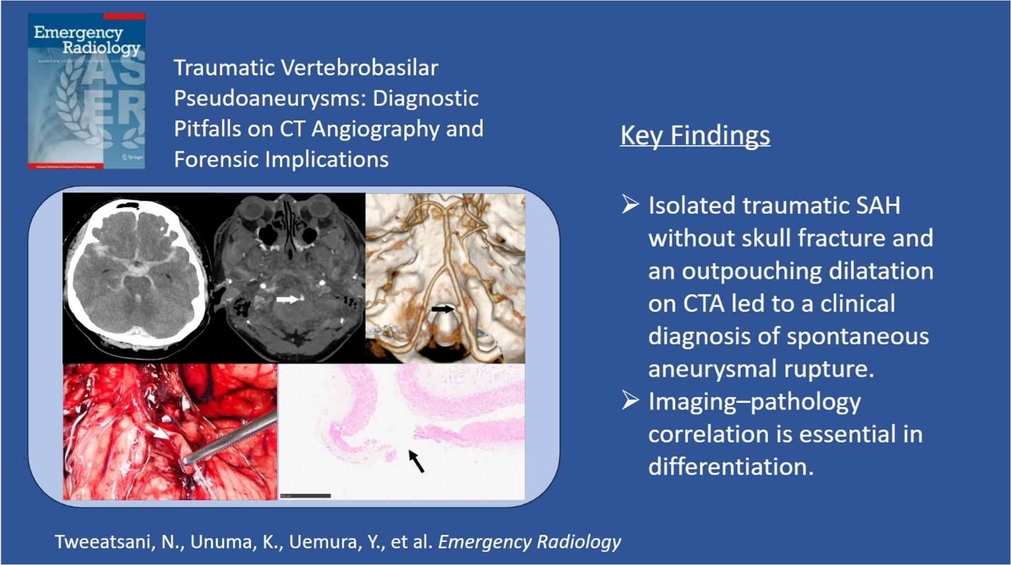
The second case emphasized the risk of misdiagnosis when imaging is interpreted without a forensic context. In this instance, basal SAH without skull fracture and an outpouching dilatation on CTA led to a clinical diagnosis of spontaneous aneurysmal rupture, and a death certificate was about to be issued as “natural death.” Autopsy findings revealed a vertical tear in the left VA, and histopathology provided definitive evidence of trauma, aligning with prior reports of trauma-induced vascular injury [6, 7]. This underscores both the absence of pathognomonic radiologic features for intracranial pseudoaneurysms [8] and the indispensable role of forensic autopsy in avoiding misclassification of cause and manner of death. Accurate distinction between traumatic pseudoaneurysms and true aneurysms is essential, as it directly impacts clinical decision-making, medicolegal classification, and attribution of responsibility. These cases demonstrate that optimized CTA protocols, cautious radiologic interpretation, and close integration with forensic pathology are crucial for reliable diagnosis in fatal traumatic SAH.
Although not applied in the present cases, postmortem CTA (PMCTA) has been proposed as a valuable adjunct when antemortem imaging is unavailable [9]. Prior studies support its utility in detecting vascular lesions that may be missed at standard autopsy, particularly in anatomically complex regions such as the skull base or posterior fossa [10, 11]. PMCTA provides a less invasive means of visualizing vascular pathology and may improve diagnostic accuracy. Future studies should evaluate its integration into forensic protocols for suspected vascular injury.



Comments (0)