
Remember me
62-year-old man with left mandibular multi-fragmented CHF following a bike accident.
Clinical FindingsSevere occlusal disturbances, ipsilateral open bite.
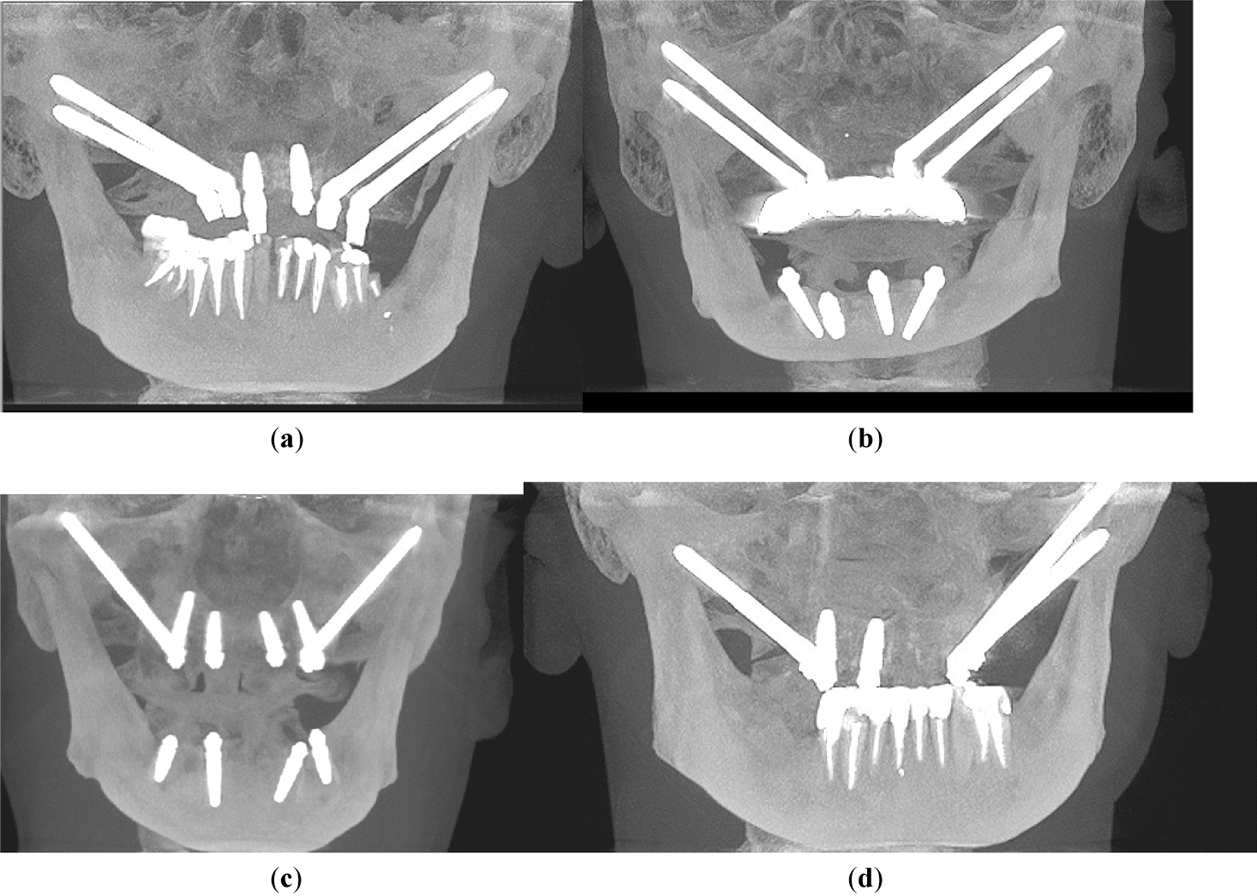
Imaging and PlanningCT-Scans delineated a left AO type-p CHF with multi-fragmentation (three major fragments), loss of vertical apposition, and anteromedial displacement (Fig. 4).
Fig. 4
Case 4: Preoperative CT-Scan (representative coronal view) – left mandibular AO type-p CHF with multi-fragmentation (three pieces) and lost vertical apposition (a). VSP/CAD – fracture reduction and PSD components (dorsal view) - large medial dome fragment (yellow), SDIMF (cyan). Dome fragment in unison with SDIMF encompassed by upper portion of primary PSD component; bipartite central window in PSD midportion; lower PSD portion surrounds postero-lateral condylar neck and base transition zone; navigation/washer PSD in its docking position; cylindrical drill guides attached (b). VSP/CAD – primary PSD component plus navigation/washer PSD in key-lock position (c). VSP/CAD – finally remaining PSD components: primary PSD removed, navigation/washer PSD in situ, T- shaped PSD over posterior condylar surface retaining SDIMF (d). Overview – STL-model (left condylar process reduced), 2 drill guides, 3 distinct PSDs (from left to right) for reduction/prefixation, navigation/washer function and fragment retention/ reinforcement (e). STL-model – dorsal view of reduced CHF with reduction/prefixation PSD component (f). Same STL-model – lateral view after tongue-in-groove docking of navigation/washer PSD and reduction/prefixation PSD (g). Same STL-model – dorso-lateral view with primary PSD removed, navigation/washer PSD left in place, additional T-shaped retaining/ reinforcement PSD (h). VSP – navigation/washer PSD placed under LSFPSO; intramedullary LSFPSO pathways (green) (i). VSP – T-shaped PSD fixated with small diameter screws (green) (j). Intraoperatively – temporary fixation of medial condylar head fragment with conventional grid plate; navigation/washer PSD fastened (k). Same view after removal of the washer and insertion of 2 lateral screws (l). Postoperative Panorex (m)
A three-component multi-purpose PSD was designed. In line with previous cases, one PSD component of the PSD was designated to reduction and prefixation. To address the difficulties in stabilizing the dislocated largest medial fragment alongside smaller pieces, the upper PSD portion was equipped with four finger-like extensions along the entire posterior surface of the condylar head. The middle section of the PSD incorporated a large window for unobstructed visualization of the fracture line and its cranial ramifications. The lower PSD portion encompassed the neck region, reaching down to the level of the deepest point of the sigmoid notch. Its anterior border contained an undulating recess serving as docking site for the second PSD component, i.e. a “tandem” washer for load distribution and reinforcement of the LSFPSO.
The upper PSD portion included six screw holes arranged in two rows (four along the common arch brace and one on each of the two central dome extensions) to ensure adequate fixation of the reassembled fragments, even if not every screw hole was occupied. The lower PSD portion contained three screw holes in a triangular configuration for anchoring to the condylar neck and base region.
The PSD “tandem” washer seated within the lateral recess along the first PSD component. This second PSD combined washer and navigation function: the tongue-in-groove design of the two components ensured accurate intraoperative transfer of the LSFPSO entry points onto the lateral fracture end at the ramus. Besides the two washer screw holes the PSD contained a third smaller hole for its temporary fixation during LSFPSO.
Detachable drill guides, designed to indicate the angulation and intramedullary pathways for the LSFPSO screws within the medial fragment assembly, were provided in two versions: two separate metallic cylinders joined by a polymer sleeve with and without a long rod-like handle.
The PSD tool set was complemented with a third component, a small T-shaped implant to augment the rigidity of the overall reconstruction after removal of the primary (reduction/prefixation) PSD. The T-shaped PSD was conceived to link the condylar neck and head and retain the small intermediate dorsal monocortical fragment (SIDMF) at the posterior condylar surface.
Surgical ProcedureThe dislocated major dome fragment, along with the smaller medial fragments, were successfully reassembled and anatomically reduced. However, repeated attempts to position the reduction/prefixation PSD component as pre-planned failed. Unresolvable collisions between the dome fragment, the cranial PSD extensions and the glenoid fossa hindered the first component from bypassing the bony crest and slipping into its planned final position.
For salvage the large medial condylar head fragment was reduced and fixated to the lateral fracture end with a conventional grid plate (1.2 mm screws; Medartis). To achieve this, it became necessary to remove the SIDMF and to close the interfragmentary gap by a slight pitch movement. This adjustment was required because the available grid plate was too short.
The entry points for the LSFPSO were established with the aid of the washer PSD. Despite the discrepancies from VSP, the placement of the washer components, drill guides and drilling could be carried out with sufficient precision. Because the bone surrounding the drill holes did not necessitate load distribution the washer PSD was removed prior to insertion of two screws (1.8 mm screws; Medartis) for the LSFPSO.
Occlusion and latero-/protrusion movements were assessed intraoperatively and confirmed satisfactory fracture reduction and stabilisation. Consequently, the grid plate was removed. Minimisation of the residual gap and SIDMF replantation were deemed unnecessary. Further stabilization with the T-shaped PSD was also not pursued.
OutcomeA postoperative panoramic X-ray confirmed anatomical reduction of the fracture.
Clinical Follow up: asymptomatic with unimpaired function.
Comments (0)