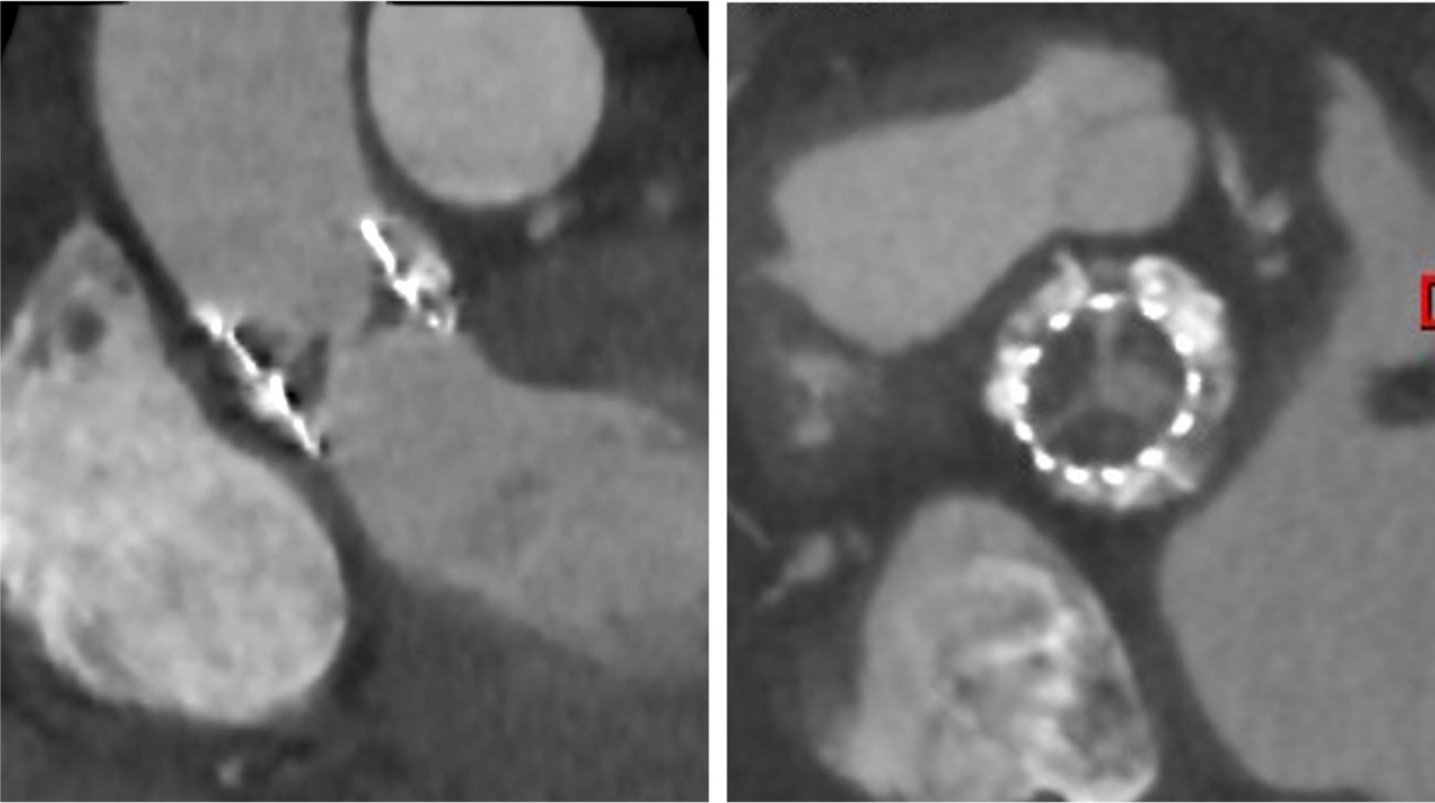
Stent apposition refers to the complete contact between the outer surface of the stent struts and the luminal contour of the vessel wall [9]. A strut is considered malapposed when the axial distance between its surface and the intima exceeds the nominal strut thickness, including any polymer coating. The clinical significance of acute malapposition remains controversial. The potential implications of malapposition are likely influenced by anatomical and procedural factors. In large-caliber vessels, minor separation may be negligible, whereas in smaller arteries or bifurcation segments, it may hinder flow, or make difficult further guidewire recrossing, increasing the risk of abluminal passage and suboptimal procedural results [3, 4]. According to the EAPCI consensus document, routine correction of acute malapposition is not recommended when the axial separation is less than 400 micrometers and the longitudinal extension is under 1 mm [9]. In such cases, spontaneous neointimal coverage is anticipated, and procedural manipulation may be avoided. Thus, while acute malapposition is not in itself an indication for further intervention, its presence should be evaluated based on vessel size, lesion complexity, and procedural goals. If needed, in the case of isolated malapposition without stent underexpansion, low-pressure inflation with a semi-compliant balloon is generally sufficient. High-pressure inflation with non-compliant balloons is usually unnecessary and may pose additional risks.
Edge Dissection
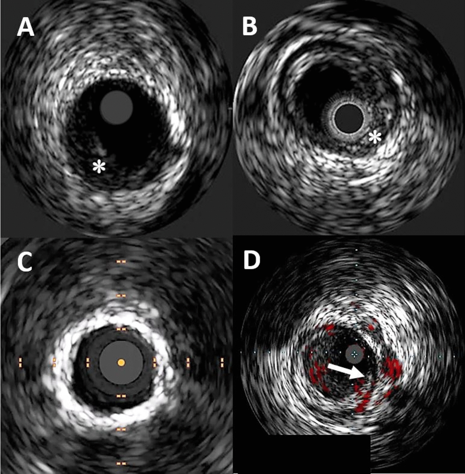
Edge dissections is defined as a tear of the luminal surface occurring at the proximal or distal stent edges [9]. These injuries may range from minor intimal flaps to deeper lesions involving medial or adventitial layers. Approximately the 80% of edge dissections are angiographically silent, and the majority heal spontaneously over time without clinical consequence [20]. Nevertheless, detailed characterization of dissection morphology, including longitudinal extension, circumferential arc, dissection depth (intimal, medial, or adventitial), flap thickness, and cavity depth (the distance between flap and the underlying plaque) is essential during results assessment, since a progression to intramural hematoma may result in acute vessel closure, or promote adverse remodeling in the long term [3]. OCT enables detailed characterization of dissection morphology, including longitudinal extension, circumferential arc, dissection depth (intimal vs. medial or adventitial), flap thickness, and cavity depth—the latter representing the distance between the flap and the underlying plaque. Although no universal criteria currently exist for defining major dissections, there is general agreement that certain morphological features confer a higher risk of adverse outcomes. While minor intimal flaps may be safely left for a conservative approach, dissections with extensive morphological features—especially when associated with medial involvement or intramural hematoma—should be treated by additional stent implantation [4, 9]. In the CLI-OPCI registry, a linear rim of tissue exceeding 200 μm in thickness was used to define a major dissection [21]. When present at the distal edge, this morphology conferred a 2.5-fold increased risk of major adverse cardiac events, including death, myocardial infarction, and target lesion revascularization. Interestingly, the same morphology did not appear to carry significant prognostic weight when observed at the proximal edge. A pre-specified analysis of the ILUMIEN IV trial demonstrated that edge dissections involving intramural hematoma with an arc ≥ 60° and a length >3 mm were independently associated with a 1.7-fold increased risk of target lesion failure, cardiac death, and target vessel myocardial infarction [18]. Notably, this elevated risk was observed even for proximal edge dissections, challenging the traditional view that distal dissections are more clinically significant. According to the EAPCI consensus document, high-risk features include a circumferential arc exceeding 60°, longitudinal extension greater than 2 mm, involvement of deep vessel layers, the presence of residual plaque burden at the dissection site, and localization at the distal stent edge [9].


Comments (0)