
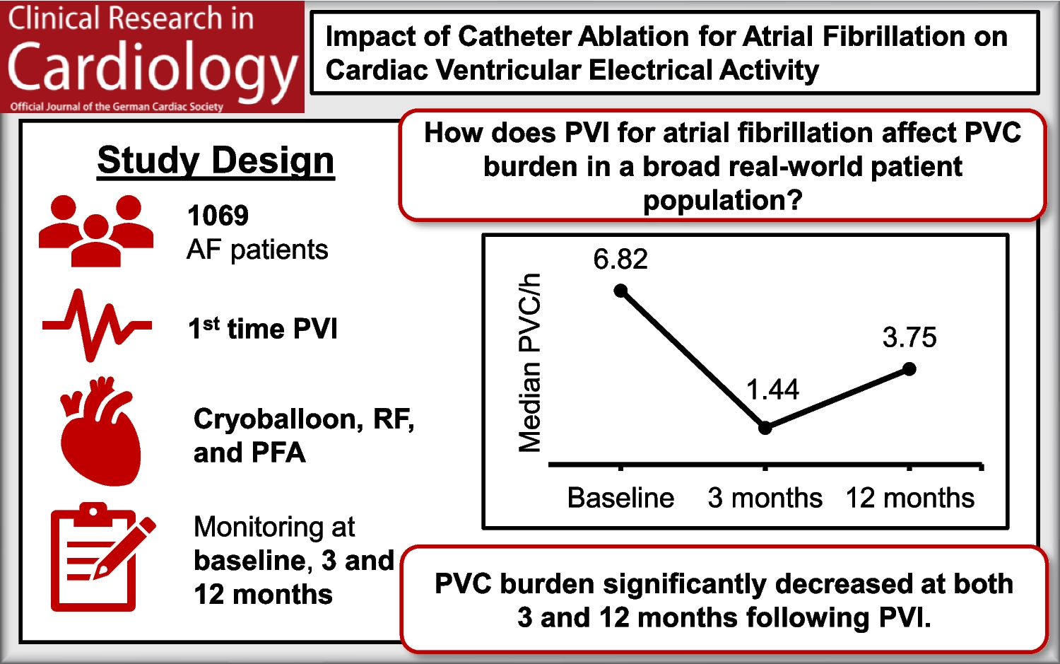
Remember me
We prospectively included 70 patients in our study. 24 (34%) underwent PFO or ASD closure, 14 (20%) underwent MitraClip implantation, 29 (41.4%) underwent pulmonary vein ablation, and 3 (4.3%) underwent left atrial appendage closure. Baseline characteristics, routine clinical laboratory values, and medication use are presented in Table 1. Compared to the primary patient cohort, PFO/ASD closure patients were younger (mean age 52.3) and a majority (87.5%) had a prior stroke considered to be due to their PFO or ASD. PFO/ASD closure patients had a lower prevalence of diabetes and atrial fibrillation. There was no significant difference in CHA2DS2-VASc score, BMI, or in sex distribution between the groups. C-reactive protein (CRP), proBNP, and D-dimers were elevated in the primary cohort (MC, PVA, and LAAC) as compared to the control cohort (PFO). Patients in the PFO cohort received more aspirin therapy and patients undergoing PVA received more factor Xa inhibition.
Table 1 Patient characteristics and risk factors for stroke. Patients undergoing catheterization with atrial transseptal puncture access were recruited and classified according to their reason for undergoing the respective procedures. History of diabetes, stroke, and atrial fibrillation were noted, along with CHA2DS2-VASc score, standard clinical laboratory parameters, and medication use. Bold font indicates statistical significance. BMI, body mass index; CRP, C-reactive protein; DAPT, dual antiplatelet therapy; LAAC, left atrial appendage closure; proBNP, pro B-type natriuretic peptideElevated NET biomarker levels in the left atriumWe hypothesized that local blood sampling would provide a more accurate picture of the thromboinflammatory state at the left atrium due to potentially disturbed blood flow associated with enlarged left atria. We therefore compared blood sampled locally from the left atrium to peripheral blood samples collected prior to the catheterization procedure from the same patients, analyzing them for both routine clinical laboratory parameters, classical markers of inflammation and thrombosis (CRP, IL-6, D-dimers, fibrinogen), as well as specialized analysis of markers of thromboinflammation (NET biomarkers, PAD4, soluble P-selectin, VWF, ADAMTS13) and cardiac damage or fibrosis (BNP, C- terminal telopeptide of type I collagen (ICTP)).
Neutrophil counts did not differ depending on the sampling site. Myeloperoxidase (MPO)-DNA complexes and citrullinated histone H3 (H3Cit) levels were significantly elevated in samples drawn from the left atrium compared to peripheral samples (Fig. 2A). Classical pro-inflammatory cytokines IL-6 and TNFα were similar in both sampling types (Fig. 2B). Although there was a modest reduction in fibrinogen in the central sample, the remainder of factors associated with a prothrombotic phenotype or endothelial activation were similar between the two types of samples (Fig. 2C). We generated correlation matrices to identify patterns in correlations in blood samples drawn through venipuncture or during catheterization directly from the left atrium (Fig. 2D). The profile is visibly similar in samples collected peripherally versus within the heart. We also performed paired analyses on the thromboinflammation parameters and confirmed similar findings as in Fig. 2A–C (Fig. 2E). As the levels of NETs were higher in the blood sample collected in the left atrium, we proceeded with all remaining analyses with these samples only.
Fig. 2
Comparison of measurements performed in peripheral venous blood or locally collected left atrial blood. A Neutrophil and NET parameters. B Pro-inflammatory cytokines. C Pro-thrombotic factors and VWF and ADAMTS13 measurements. D Spearman correlation matrices for measurements performed in peripheral blood samples (left) or central LA blood samples (right). E NET-related measurements in paired comparisons between peripheral and central blood samples. MPO-DNA, myeloperoxidase-DNA complexes; H3Cit, citrullinated histone H3; PAD4, peptidylarginine deiminase 4; IL-6, interleukin-6; TNF-⍺, tumor necrosis factor- ⍺; sPsel, soluble P-selectin; VWF, von Willebrand factor; ADAMTS13, a disintegrin and metalloproteinase with a thrombospondin type 1 motif, member 13; Ag, antigen; Act, activity; LA, left atrium. Mann–Whitney U-tests were used to determine significance in A–C and Wilcoxon tests for data in E. *P < 0.05, **P < 0.01, ****P < 0.0001; ns, not significant. N = 70
Distinct thromboinflammatory profiles based on procedure typeWe next analyzed the same parameters according to the type of procedure that was performed, combining MitraClip and LAAC patients into a combined cohort based on similar clinical characteristics (high presence of atrial fibrillation, NTproBNP, CRP, and CHA2DS2-VASc score). Here, neutrophil counts were highest in the MC + LAAC cohort as compared to PFO or PVA but there was no difference in levels of NETs with the exception of higher H3Cit in MC + LAAC compared to PFO patients (Figure S1A). MC + LAAC and PVA had higher IL-6 compared to PFO, whereas TNF-alpha was elevated only in MC + LAAC compared to PFO patients (Figure S1B). Notably, fibrinogen was the only pro-thrombotic marker that was not elevated in MC + LAAC patients (Figure S1C). Von Willebrand factor activity and antigen were highly elevated in MC + LAAC patients with a mean of twice normal-range values. Accordingly, ADAMTS13 activity and antigen levels were reduced in MC + LAAC but not in PVA patients, compared to the PFO cohort.
Analysis of troponin T, proBNP, and ICTP confirmed a higher degree of cardiac damage or fibrotic remodeling in MC + LAAC patients, with PVA patients having only elevated troponin T or proBNP compared to PFO patients (Figure S1D). Furthermore, echocardiographic measurements of body surface area corrected- left atrial size (left atrial volume index, LAVI) or LA flow velocity, as well as ejection fraction and E/E’ ratio were analyzed for each patient. MC + LAAC patients had reduced LA flow velocity, enlarged left atria, higher E/E’ compared to both PVA and PFO cohorts, and lower EF compared only to the PFO cohort (Figure S1E).
Spearman correlation coefficients were calculated for the different parameters measured above, with correlation matrices shown in Figure S2. In contrast to the sampling type, there was a clear difference in the correlation matrix depending on the procedure type. Heat maps showed distinct profiles among the patient groups with more significantly positive correlations between thrombotic markers, cardiac damage biomarkers, and NET measurements were seen in the MC + LAAC group (Figure S1F). Finally, a principal component analysis was performed with all study measurements described above to show the clustering the individual patients according to procedure type (Figure S1G). This analysis showed that the PFO group clustered distinctly from the others, with less variation in parameters. The greatest degree of variation in patient profile distribution was evident in the MC + LAAC cohort, with PVA patients clustering between the two. The biplot which shows the contribution of each measured parameter to the PCA is shown in Figure S3. In summary, we could identify differences in the measured blood parameters depending on the procedure type, along with expected differences in cardiac parameters using biomarkers in blood or imaging.
Chronicity of atrial fibrillation correlates with thromboinflammatory profileFinally, we performed similar analyses by grouping patients according to the presence or absence of atrial fibrillation (AF). Similar results were obtained to the LAVI cutoff stratification, with an additional significant increase in H3Cit, IL-6, D-dimers, soluble P-selectin, and ICTP in patients with any history of atrial fibrillation (Fig. 3A–D). The ejection fraction was also significantly reduced in the AF group (Fig. 3E). The heterogeneity across the AF patients and the PCA plots was similar to what was seen in patients with enlarged LAVI (Fig. 3F, G). Correlation matrices for all parameters measured for patients in SR and any AF are depicted in Figure S4. We also confirmed similar findings in the peripheral blood samples collected by traditional venipuncture (Figure S5).
Fig. 3
Comparison of left atrial blood measurements and cardiac parameters in patients with sinus rhythm (SR) or atrial fibrillation (AF). A Neutrophil and NET parameters. B Pro-inflammatory cytokines. C Pro-thrombotic factors and VWF and ADAMTS13 measurements. D Biomarkers of cardiac damage/remodeling. E Structural and functional measurements as performed by echocardiography. F Heat map showing reduced dimensionality data with relative Z-scores for each parameter. G Principal component analysis (PCA) showing clustering of procedure groups by variance. MPO-DNA, myeloperoxidase-DNA complexes; H3Cit, citrullinated histone H3; PAD4, peptidylarginine deiminase 4; IL-6, interleukin-6; TNF-⍺, tumor necrosis factor- ⍺; sPsel, soluble P-selectin; VWF, von Willebrand factor; ADAMTS13, a disintegrin and metalloproteinase with a thrombospondin type 1 motif, member 13; Ag, antigen; Act, activity; LA, left atrium; CK-MB, creatine kinase MB; proBNP, brain natriuretic peptide; ICTP, C-telopeptide of collagen I; LAA, left atrial appendage; LAVI, left atrial volume index; EF, ejection fraction. SR, N = 27; AF, n = 43. Mann–Whitney U-tests were used to determine significance. *P < 0.05, **P < 0.01, ***P < 0.001; ****P < 0.0001; ns, not significant
In parallel, we also performed a different subgroup analysis where we classified the AF patients based on permanent AF status and including the intermittent AF patients together with those who are in sinus rhythm, as at the time of blood sampling these patients were also in sinus rhythm (SR) (Figure S6). In contrast to the classification with all AF patients, with examination of permanent AF only lost the distinct profile based on our measured parameters and several of the significant differences between the AF and SR, including VWF/ADAMTS13 measurements. This indicates that a prior history of AF episodes rather than active AF at the time of sampling is more predictive of thromboinflammation.
Correlations between NETs, VWF/ADAMTS13 axis, and cardiac injury with LA sizeIndividual Spearman correlation plots are shown in Fig. 4. Neutrophil numbers and H3Cit levels correlated positively with LAVI, proBNP, and ICTP (Fig. 4A, B), indicating a link with fibrosis development and ongoing cardiac damage. VWF antigen was also significantly positively correlated with the same factors, with ADAMTS13 activity conversely negatively correlated (Fig. 4C, D). PAD4 levels correlated with reduced ADAMTS13 activity levels correlated with higher values throughout all NET markers in LA blood (Fig. 1D), and PAD4, which has been shown to render ADAMTS13 inactive [22]. This suggests a potential role for PAD4-mediated citrullination of ADAMTS13 in its reduced activity, in addition to lower levels of circulating antigen.
Fig. 4
Spearman correlation analyses of left ventricular volume index (LAVI) and cardiac injury biomarkers with selected thromboinflammation parameters. Neutrophil counts (A), citrullinated histones (B), and VWF antigen (C) positively correlated with LAVI, proBNP, and ICTP. Conversely, ADAMTS13 activity (D) negatively correlated with LAVI, proBNP, and ICTP. H3Cit, citrullinated histone H3; VWF, von Willebrand factor; ADAMTS13, a disintegrin and metalloproteinase with a thrombospondin type 1 motif, member 13; Ag, antigen; Act, activity; r, Spearman correlation coefficient. N = 70
Enlarged LAVI correlates with a distinct thromboinflammatory profileIn parallel, echocardiographic measurements including left atrium size and flow velocity, as well as ejection fraction of the left ventricle and E/E’ were recorded and analyzed. A cutoff for enlarged left atria using a left atrial volume index of greater than 34 was used according to European Society on Cardiology guidelines [10]. Here, neutrophil numbers, H3Cit, TNFα, and IL-6, but not MPO-DNA, were elevated in the enlarged LAVI subgroup (Fig. 5A, 5B). In the thrombotic factor measurements, once again VWF activity and antigen were both highly elevated, with a concurrent reduction in ADAMTS13 activity and antigen (Fig. 5C). The cardiac damage markers were higher in those patients with large LAVI, and LA flow velocity was reduced while E/E’ was increased in the patient cohort (Fig. 5D, E). We also performed correlation analyses with the measured biochemical parameters in blood and found that there was heterogeneity in the profiles across the different donors (Fig. 5F). The PCA also identified differences in the profiles of the normal vs larger LA samples (Fig. 5G). As the PFO cohort had significantly smaller LAVI, we also performed a subgroup analysis without the inclusion of these patients. Here, we also saw a similar increase in neutrophils, IL-6, VWF, and a subsequent decrease in ADAMTS13 parameters (Figure S7).
Fig. 5
Comparison of left atrial blood measurements and cardiac parameters in patients with normal versus enlarged left atria (LA). Patients were stratified by LAVI with 34 as a cutoff for enlarged LA. A Neutrophil and NET parameters. B Pro-inflammatory cytokines. C Pro-thrombotic factors and VWF and ADAMTS13 measurements. D Biomarkers of cardiac damage/remodeling. E Structural and functional measurements as performed by echocardiography. F Heat map showing reduced dimensionality data with relative Z-scores for each parameter. G Principal component analysis showing clustering of procedure groups by variance. MPO-DNA, myeloperoxidase-DNA complexes; H3Cit, citrullinated histone H3; PAD4, peptidylarginine deiminase 4; IL-6, interleukin-6; TNF-⍺, tumor necrosis factor- ⍺; sPsel, soluble P-selectin; VWF, von Willebrand factor; ADAMTS13, a disintegrin and metalloproteinase with a thrombospondin type 1 motif, member 13; Ag, antigen; Act, activity; LA, left atrium; CK-MB, creatine kinase MB; proBNP, brain natriuretic peptide; ICTP, C-telopeptide of collagen I; LAA, left atrial appendage; LAVI, left atrial volume index; EF, ejection fraction. Normal atria (N), n = 37; enlarged atria (E), n = 33. Mann–Whitney U-tests were used to determine significance. *P < 0.05, **P < 0.01, ***P < 0.001; ****P < 0.0001; ns, not significant. N = 70
In univariable linear regression analyses, age (β, 0.39; CI, 0.37–1.14, p < 0.001), neutrophils (β, 0.39; CI, 0.17–0.64, p = 0.001), VWF Ag (β, 0.34; CI, 0.12–0.64, p = 0.004), and H3Cit (β, 0.31; CI, 0.03–0.20, p = 0.01) were associated with LAVI. Added to a multivariable model with LAVI as dependent variable, age (β; 0.26; CI, 0.004–0.95, p = 0.048) and neutrophil count (β; 0.32; CI, 0.06–0.63, p = 0.017) remained associated with LAVI, but VWF Ag (β; 0.065; CI, − 0.24–0.38, p = 0.65) and H3Cit (β; 0.068; CI, − 0.07–0.12, p = 0.59) were no longer associated with LAVI. In a subgroup analysis with the same multivariate analysis without the PFO cohort, only neutrophil count remained significant (Table S1). In a multiple regression analysis with NETs as the dependent variables, neutrophil counts (p = 0.0493), PAD4 (p = 0.0066), D-dimer (p = 0.0142), VWF antigen (p < 0.001), proBNP (p = 0.0322), and E/E’ (p = 0.0491) independently associated with H3Cit whereas they did not associate with MPO-DNA.
Finally, we followed up with patients over the 12 months following initial inclusion in the study to identify thrombotic events or major bleeding episodes. Two patients died in the year after their procedure. One patient suffered from a femoral fracture and acute kidney failure, whereas the second had progressive dysphagia of multifactorial origin including an acute ischemic stroke. Two patients suffered an acute ischemic stroke, and eight patients suffered from bleeding events. The low number of events precluded further analysis within the study design.
Comments (0)