
Remember me


This study was a single-blinded randomized six-month clinical trial (RCT) approved by the National Ethics Review Board in Uppsala, Sweden (2020-02586). The RCT was conducted according to the World Medical Association Declaration of Helsinki principles, revised in 2013. The CONSORT guidelines for clinical trials were followed. The study was registered under www.clinicaltrials.gov (study no. NCT05772299).
Study populationThe patients were referred for a treatment of peri-implant mucositis. Sixty-four patients were screened and reviewed for medical history and available radiographs. Potential cases, according to the inclusion and exclusion criteria, further underwent a full mouth routine periodontal examination. Forty-six subjects who fulfilled the inclusion criteria and consented were consecutively included.
Inclusion criteria—presence of peri-implant mucositisOne dental implant with a minimum of one or more peri-implant sites with probing depth ≥ 4 mm combined with bleeding and/or pus on probing
A limited allowed bone loss ≤ 2 mm measured from the implant shoulder (changes resulting from initial bone remodelling)
Exclusion criteriaSubjects with uncontrolled diabetes mellitus (HbA1c > 6.5)
Subjects requiring prophylactic antibiotics
Subjects taking prednisolone
Subjects taking medications with effects on gingival growth
Subjects taking systemic antibiotics in the preceding month
For confidentiality, coding was used and saved according to the regulations of Kristianstad University, Sweden. The patients were referred for treatment between 2021 and 2023. Due to Covid 19, when no patient treatment was performed the start of the study was delayed. Before entering the study, a preparatory treatment phase was performed, including instruction in oral hygiene measures and debridement of teeth and implants not to be part of the study. Good patient motivation and compliance were also identified. The patients were informed about the study and filled out written consent.
RandomizationAfter entering the study, patients were randomly assigned to treatment with an Er:YAG laser (test) or an ultrasonic device (control) using pre-prepared randomization according to pre-defined computerized randomization.
MeasurementsThe clinical study was conducted at the speciality clinic of periodontology at Kristianstad University. Unaware of the treatment, the same examiner (CLL) performed the measurements, and the patients were asked not to talk about their treatment. Measurements were recorded at baseline and in months one, three, and six using a manual plastic probe tip diameter of 0.5 mm (Colorvue Probe, Hu-Friedy, USA) and probing force of 0.2 N., The following measurements were recorded for the full mouth: bleeding on probing (FMBoP) (4 sites/tooth, 6 sites/implant) [11], plaque score (FMPS) (4 sites/tooth, 6 sites/implant) [12]. Measurements at the implant were: bleeding on probing (BoP) (6 sites), severity of bleeding graded 0–3 0 = no bleeding, 1 = a dot of bleeding, 2 = a line of bleeding, and 3 = a drop of bleeding (6 sites) (i.e. modified bleeding index or mBI) [5, 13], suppuration (6 sites), plaque (6 sites), probing pocket depths (PPDs) (6 sites), hyperplasia, buccal recession of the mucosal margin.
Patient evaluation of the treatment: Oral health–related quality of life (OHQoL) was measured with the Oral Health Impact Profile–14 version (OHIP-14) [14] at baseline and month six. The Quality of life was expressed as the sum of the fourteen questions, a maximum of 56 scores. The mean values were calculated. A lower score indicates high OHQoL, and a higher score indicates low OHQoL. The analysis did not include patients who had not answered all questions.
Visual Analogue Scales consisted of a 10-point graded straight horizontal line, with the left end indicating 0 "no pain and the right end indicating 10 "worst pain". The VAS concerned pain during treatment, the same day, day two and day three after the treatment. Further, the other VAS reflected the patients' aesthetic satisfaction at baseline and month six. The left end on the VAS indicated "not at all satisfied", and the right was "very satisfied". Each patient's mean visual analogue score was calculated.
Radiographic examinationIntraoral digital periapical standardized radiographs of the site of interest were taken with a bite-block to detect loss of bone at baseline and month six. Radiographs were analysed by an independent and blinded examiner (VWB) using the Synedra View software for personal computers. The known distance between implant thread pitch lengths was used to calibrate the images. The distance between the implant shoulder and the most superior alveolar bone level on the mesial and distal aspects of the implant was measured and merged. The intra-class coefficient (ICC) for the distance between the implant shoulder and the alveolar bone level was 0.94 (95% CI: 0.84–0.98, p < 0.00).
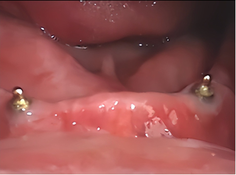
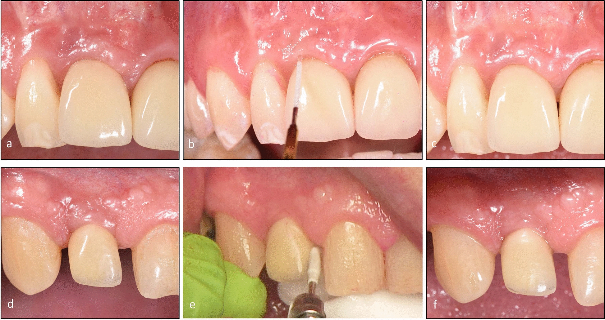
Treatment proceduresThe same treatments were performed at baseline, months three and six. Final evaluation data was provided in month six. All treatments were performed by a non-blinded periodontist (VWN). No implant-supported constructions were removed, and no local anaesthesia was used. At each session and after plaque registration, the patients were instructed to use proper home care using a toothbrush and interproximal aids as needed. Treatment in the test group (test) was done using an Er: YAG laser (Erbium-doped yttrium–aluminium-garnet; Er: YAG, AdvErL EVO, Morita Corporation, Japan—> J. MORITA MFG. CORP., Japan) with tip PS400TS and power 50 mJ, frequency 10 PPS and proportion air/water 7/7 [15]. The tip was placed in the peri-implant pocket, and a moving motion was from bottom to coronal at the implant surface, with a sweeping motion covering the soft tissue. The treatment in the control group (control) was performed with an ultrasonic device (Electro Medical Systems, EMS) with a plastic-coated tip (PI Instrument Nyon; EMS, Nyon, Switzerland). The tip was placed in the peri-implant pocket and used in a similar way as described above (Fig. 1). All treatments were finalized by polishing with a rubber cup and polishing paste. Postoperative complications were asked for in the examination protocol as an open question at each examination, and treatment time was registered.
Fig. 1
Peri-implant mucositis before, during and after treatment with Er: YAG laser or ultrasonic scaler. a Peri-implant mucositis 11 before treatment b Er: YAG laser treatment 11 c six months after Er: YAG laser treatment 11 d Peri-implant mucositis 12 before treatment e ultrasonic scaling 12 f six months after ultrasonic scaling 12
At month one, measurements, oral hygiene instructions, and polishing with a rubber cup and polishing paste were performed (CLL).
Statistical analysisThe IBM SPSS version 27.0 statistical software package (SPSS Inc., Armonk, NY, USA) for personal computers was used in the analyses. The statistical unit was based on both the patient and the implant. Descriptive and comparative statistics were used when analysing the data. The primary outcome measure in this study was bleeding on probing (BoP). A power analysis was performed, and thirty-eight patients were minimum to reach a clinically significant difference at ± 30% reduction in bleeding of probing (BoP) between the groups with alpha 0.05 and power 80%. Forty-six individuals were included in the study, and all analyses were based on the "per protocol" principle. Mean values, standard deviation (SD) and frequency distributions are given. Independent t-test (equal variance not assumed) and paired t-test were used to compare inter- and intra-group differences. The chi-square test and non-parametric McNemar test were used for categorical variables. Statistical significance was set at p < 0.05.
Comments (0)