
Remember me

In three female cases (Figs. 1 and 2) there was found a peculiar anatomic variation of the sigmoid sulcus and sinus: an inferior recess of the sulcus was filled by an inferior diverticulum of the sigmoid sinus and projected inferiorly from the jugular process of the occipital bone. This leads to a 6% general and 12% prevalence in females.
Due to this topography, that diverticulum was termed „paramastoid diverticulum of the sigmoid sinus” (PMDSS). The variant was unilateral in all the cases. In cases #1, #2 and #3, the heights (measured from the bottom of the sigmoid sulcus) and inner diameters of the PMDSSs were, respectively, 9.94/11.01 mm, 8.21/4.85 mm and 5.97/8.72 mm. In each of these cases, a high jugular bulb was also found on the side with PMDSS (Fig. 2). The high jugular bulb in cases #1 and #3 had superior diverticula. The bony bottom of each PMDSS was extremely thin.
In case #1, the lateral condylar vein crossed the PMDSS medially to join the plexus of the vertebral artery infero-medially to the PMDSS. The tip of that PMDSS reached 3.48 mm superior to the transverse process of the atlas. In case #2, the lateral condylar vein also coursed medially to the PMDSS through a deep paracondylar groove between the occipital condyle and the PMDSS (Fig. 3A).
In cases #1 and #2, the occipital artery coursed beneath the occipitomastoid fissure on the lateral side of the PMDSS.
In case #3, the tip of the PMDSS herniated through the bottom of the bony diverticulum to contact extracranially the posterior condylar vein on that side (Fig. 3B and C). The lateral condylar vein laterally surrounded the PMDSS and joined the posterior condylar vein suboccipitally. The occipital artery coursed laterally to both the PMDSS and lateral condylar vein.
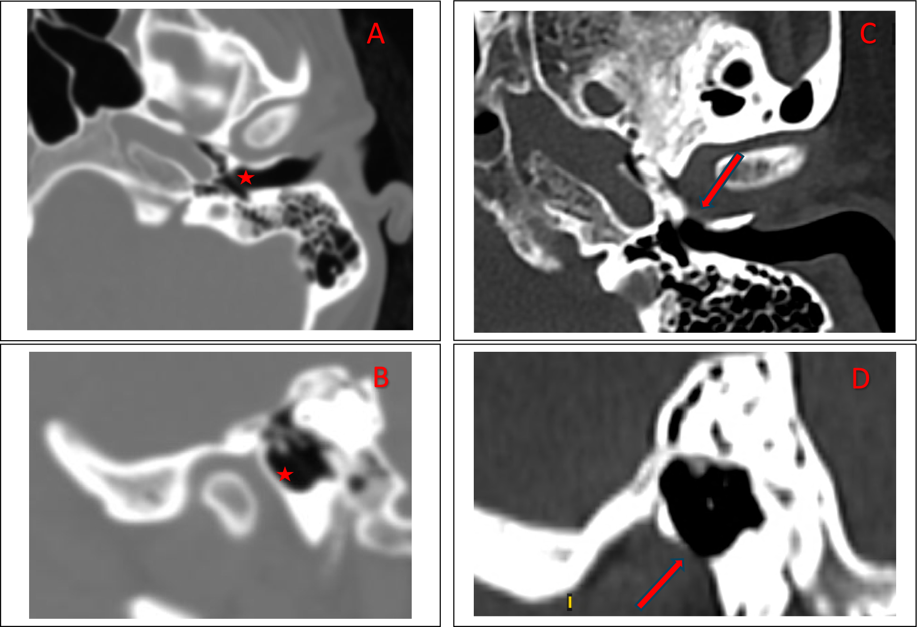
Fig. 1
Correlated two-dimensional slices in the anatomical planes, sagittal (A, D, G), axial (B, E, H) and coronal (C, F, I) in the three cases with a paramastoid inferior diverticulum of the sigmoid sinus. The axial sections are viewed inferiorly, and the coronal sections are viewed anteriorly. The anatomical variation is on the right side in cases #1 (A, B, C) and #2 (D, E, F). The variant in case #3 (G, H, I) is on the left side. (1) internal jugular vein; (2) paramastoid diverticulum of the sigmoid sinus; (3) transverse process of the atlas; (4) mastoid process
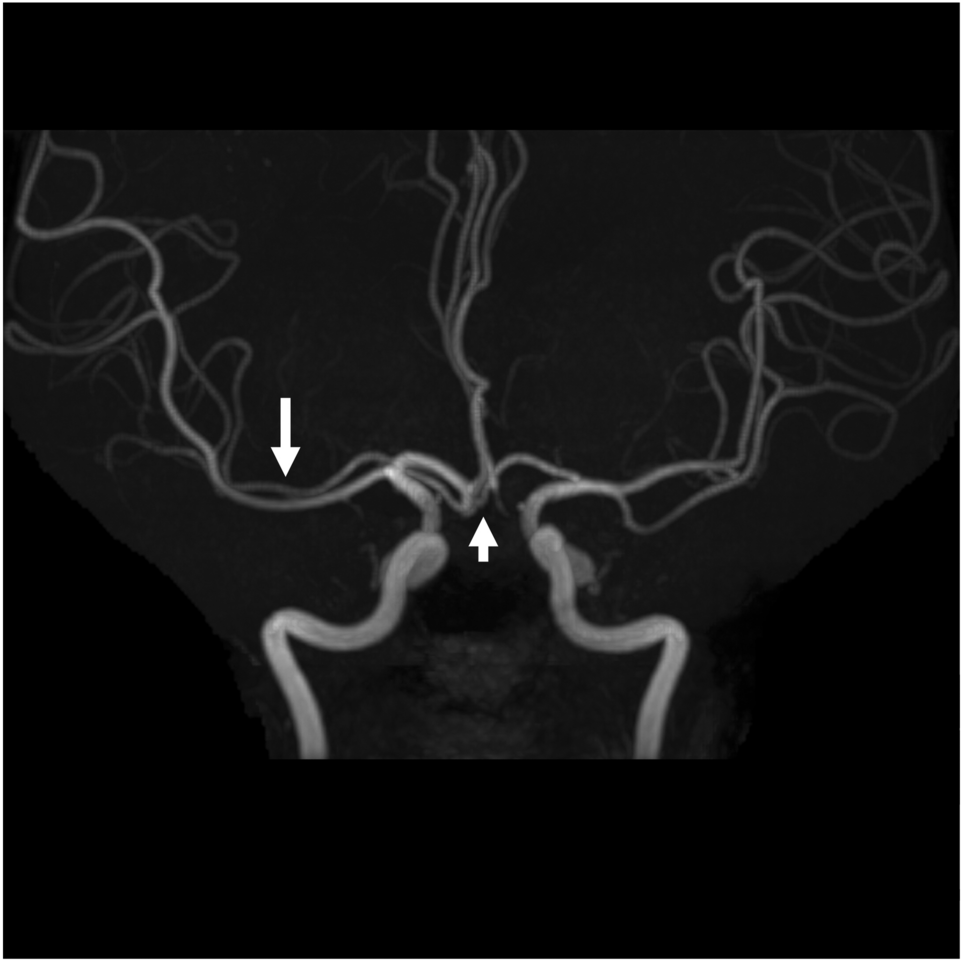
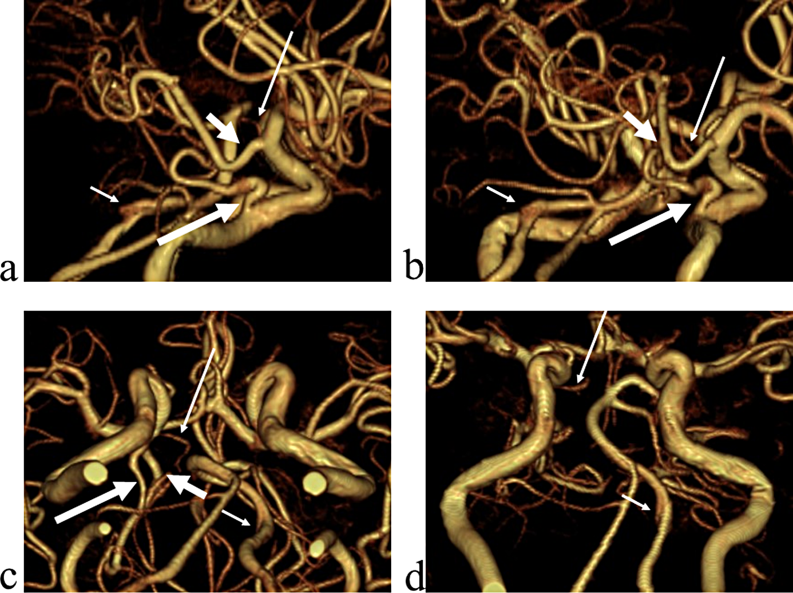
Fig. 2
Three-dimensional volume renderings of the inferior paramastoid diverticula of the sigmoid sinuses in cases #1 (A, right side, lateral view), #2 (B, right side, lateral view) and #3 (C, left side, lateral view). (1) sigmoid sinus; (2) internal jugular vein; (3) jugular bulb; (4) superior diverticulum of the jugular bulb; (5) inferior paramastoid diverticulum of the sigmoid sinus
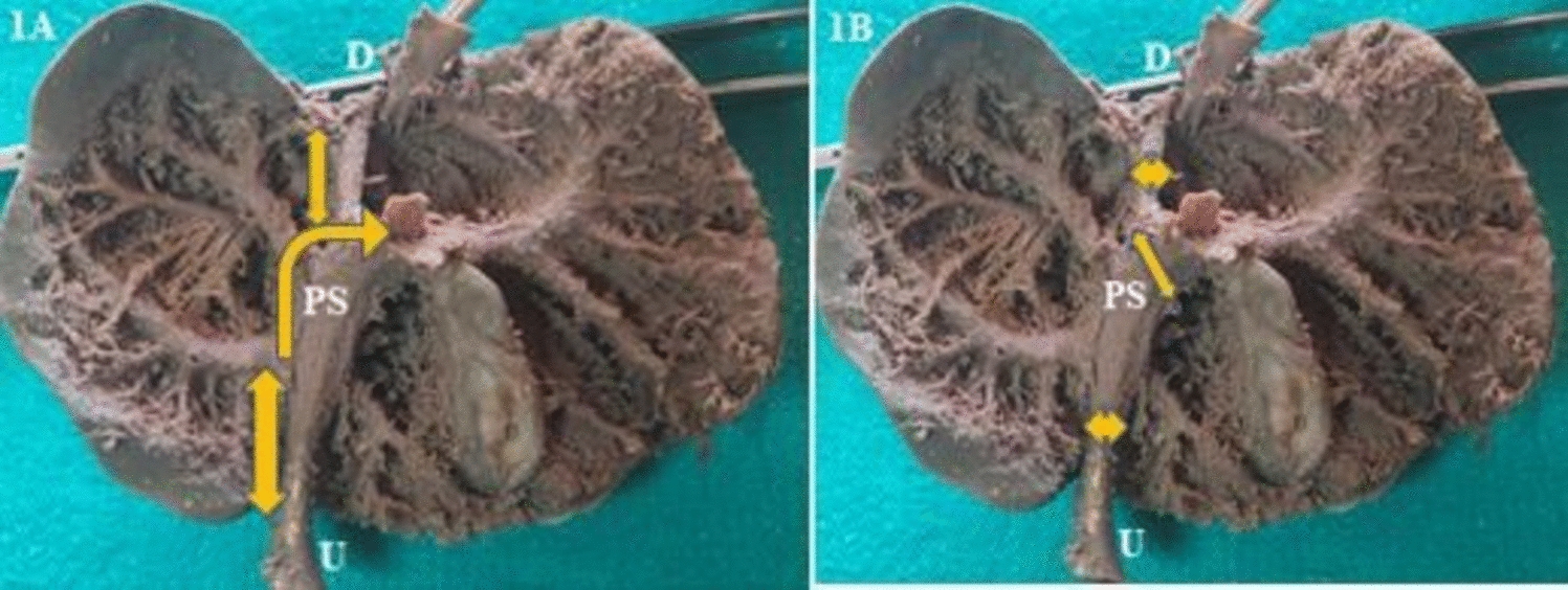
Fig. 3
A. Coronal section through the paramastoid diverticulum of the right sigmoid sinus (PMDSS) in case #1. Anterior view. (1) transverse process of the atlas; (2) PMDSS; (3) paracondylar groove; (4) occipital condyle; (5) lateral condylar vein; (6) venous plexus of the vertebral artery. B, C. Successive axial sections through the PMDSS in case #3. Inferior views. (1) tip of the PMDSS; (2) posterior condylar vein; (3) lateral condylar vein; (4) internal carotid artery; (5) internal jugular vein; (6) styloid process; (7) occipital artery
Comments (0)