Infertility, is a serious human reproductive health problem, affecting about 15 % of the couples of childbearing age worldwide [1]. Male factors account for about 50 % of infertile couples [2], and can be caused by various adverse factors, such as genetic problems, environmental factors, and chemotherapy drugs [[3], [4], [5]]. Previous investigations have shown that chemotherapy drugs for cancer patients often impair the physiological function of the male reproductive system, particularly alkylating chemicals generating the most visible sperm destruction [6]. High doses of alkylating chemicals can cause persistent or permanent azoospermia, which is hazardous to male fertility [6,7]. Busulfan, an alkylating agent, is commonly used for the treatment of chronic myelogenous leukemia [8] and for the preparation of hematopoietic stem cell transplantation [9]. However, long-term use of busulfan can result in bone marrow suppression [10], pulmonary interstitial fibrosis [11], decreased fertility, and even infertility in patients [6]. According to researches, the main target cells of busulfan-causing male reproductive problems are spermatogonia [12,13], which is a group of stem cells located on the spermatogenic epithelium of testis, and is the basis of maintaining spermatogenesis and male fertility. Once spermatogonia is damaged, it will cause spermatogenesis disorders, or even lead to oligospermia and azoospermia. Therefore, clarifying the molecular mechanisms of busulfan-induced spermatogonial damage could provide a theoretical basis for understanding busulfan-induced male reproductive toxicity and even provide potential targets for clinical intervention in oligospermia.
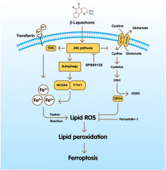
Ferroptosis is a novel regulated cell death type, typical biochemical features of which are iron-dependent lipid peroxidation and inactive of Xc-GSH-GPX4 antioxidant systems [14]. Growing evidence displayed that some male reproductive disorders directly result from ferroptosis caused by the accumulation of iron and lipid peroxides in testis, which can cause damage to sperm DNA, testosterone synthesis, and blood-testis barrier [15]. And inhibition of ferroptosis by lessening the formation of lipid peroxidation products or intracellular iron accumulation can partly alleviate multifactorial-induced male reproductive disorders [16]. It follows that ferroptosis may be a promising therapeutic target for the male reproductive system diseases. Importantly, ferroptosis has recently been implicated in busulfan-induced oligospermia in mice [17]. Nevertheless, whether spermatogonia ferroptosis is involved in busulfan-induced oligospermia and the specific molecular mechanisms remain unclear.
Autophagy is a biochemical process that degrades cytoplasmic components, proteins, macromolecules, and aberrant organelles via lysosomal dependent pathways [18]. The process is essential for cell survival and maintenance; nevertheless, excessive or deficient autophagy can result in cell death [19]. Recent researches have shown that autophagy participates in regulating ferroptosis through the degradation of ferritin, which is mediated by nuclear receptor coactivator 4 (NCOA4) via binding to ferritin heavy chain 1 (FTH1) [20]. The complete process, called ferritinophagy, is involved in the regulation of iron release and is required for intracellular iron bioavailability [21]. Under the physiological state, when the intracellular iron content is low, ferritin is carried to the autophagosome by NCOA4 and degraded in the lysosome, therefore boosting the intracellular free iron level [22]. Moreover, excessive ferritinophagy causes intracellular iron accumulation and ferroptosis [23]. Previous researches found that autophagy participates in busulfan-induced spermatogenesis disorders and oligospermia, but the specific role is still controversial [[24], [25], [26]], and whether it mediates the occurrence of spermatogonial ferroptosis caused by busulfan is unclear.
Based on the above questions, we speculated that busulfan could mediate ferroptosis in spermatogonia through autophagy-dependent ferritin degradation, and subsequently promote azoospermia. To verify the hypothesis, firstly, GC-1 spg cells were treated with busulfan, Fer-1, DFO, and/or 3-MA, and then cell death, ferroptosis and ferrritinophagy were detected. Thereafter, mice were exposed to busulfan and/or 3-MA, and ferroptosis, ferrritinophagy, testis morphology as well as spermatogenesis were detected. These results explain the toxicity mechanisms of busulfan on the male reproductive system and identify a new target for the therapy of busulfan-induced male infertility.


Comments (0)