
Remember me

All patients born between 2002 and 2013 (aged between 6 and 18 years) diagnosed with an AWD and treated at our Department were invited to participate in a prospective study consisting of clinical examination, spirometry, cardiopulmonary exercise performance testing (CPET), assessment of the motor activity, ultrasound and electromyography of the abdominal wall, stance and gait analyses and questionnaires for gastrointestinal QOL. All measurements were performed in one day. Patients with hemodynamic relevant cardiac disease or mental disorders were excluded.
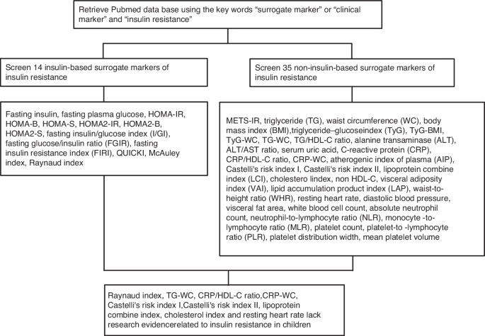
According to the literature, we classified GS as complicated if patients suffered from concomitant intestinal atresia, volvulus, necrosis or perforation.16,17,18 Giant OC was defined as a defect larger than 5 cm.19,20
The results were compared to a healthy age-, sex-, BMI- and physical activity level-matched control group recruited from friends and family of the Department´s employees.
This study was performed according to the declaration of Helsinki. All patients and controls and/or their legal guardians gave informed written consent. This study was approved by the institutional review board (EK 32–231 ex 19/20). All measurements were performed between May 2020 and June 2021.
Anthropometric dataBody height (BH) and weight (BW) were measured and the body mass index (BMI) calculated. Segmental multi-frequency impedance spectroscopy (CombynTM ECG, Academic Technologies at the Institute of Cardiovascular Medicine GmbH, Graz, Austria) was used to measure appendicular muscle mass and total body fat (TBF) as previously described in the literature.21 Cardiac arrhythmias were excluded with a 12-lead resting electrocardiography (ECG) and non-invasive blood pressure (NIBP) measurement at rest was performed.
Participants were asked to rate their physical activity levels according to four groups (“daily”, “several times a week”, “once per week” or “once per month”).
SpirometryLung function was measured by small spirometry (Oxycon Pro® Carl Reiner GmbH, Vienna, Austria) at rest and following exercise. Maximum vital capacity (VCmax) and the forced expiratory volume in 1 s (FEV 1) were assessed.
VCmax was expressed as observed and corrected according to the expected maximum vital capacity over age and sex. The Tiffeneau index was calculated as FEV 1/VCmax. A restrictive ventilation disorder was defined as a predominantly decreased VCmax and an obstructive ventilation disorder as a decreased Tiffeneau index.22
Cardiopulmonary exercise performance testing (CPET)CPET with a bicycle ergometer (Excalibur Sport®, Lode B.V., Groningen, The Netherlands) and the spirometer in an upright position was used to measure cardiopulmonary exercise performance. A stepwise load increase protocol, specified for sex and age, was used as published before.23 The spiroergometry was continued to subjective exhaustion or until the participants were unable to maintain the required pedaling speed (cadence) of more than 60 revolutions per minute (rpm). A three minutes “cool down” of slow pedaling (60 rpm) with the same workload as at the beginning of the test followed the exercise phase.
Twelve-lead ECG (Cardinal HealthTM electrocardiography, Dublin, Ireland) measured Heartrate (HR) and finger pulse oximeter (Habel Medizintechnik®, Vienna, Austria) assessed oxygen saturation continuously during the whole exercise.
At the end of each step and after the cool down lactate levels were determined by earlobe sampling of 20 µl blood per measurement to heparinized capillaries before the test (enzymatically amperometric measurement with a Biosen C_line® (EKF Diagnostics for life, Cardiff, UK)).
Respiratory parameters including the oxygen uptake (VO2), the oxygen pulse (O2/HR), the respiratory equivalent for oxygen (EQO2), the breathing reserve (BR) and the respiratory exchange ratio (RER) were assessed.23
Relative performance capacity was calculated from the achieved maximal wattage in relation to age and sex-specific standard values.24 The peak oxygen uptake (peak VO2) was defined as the average VO2 over the last 30 seconds prior to subjective exhaustion and was expressed in ml/kg/min. A RER > 1.10 was used as criterion to determine that the peak VO2 reflects a peak physiological workload.25
Assessment of motor abilitiesThe Dordel-Koch-Test (DKT) was used to assess motor abilities (flexibility, coordinative and conditional skills).26 The tests consists of seven established and validated items: lateral jumping, sit and reach, sit-ups, long stand jump, one-legged stand, push-ups and 6-min-run and allows a quick and differentiated evaluation of motor performance among all basic motor skills.26 In the present study, the endurance was tested with a spiroergometry instead of a 6-min-run. The indicated grades 1 to 6 correspond to a school grading system with lower values indicating better performance.26
Electromyography (EMG) of the abdominal wall & gait and stance analysisEight sensors as shown in Supplementary Fig. 1 were fixed to the abdominal wall (Ultium® Wireless Surface EMG, Velamed GmbH with Ultium® EMG Sensor, Velamed GmbH and Noraxon MR 3.14, Cologne, Germany). Afterwards, patients had to perform eight exercises to measure activity of the M. rectus abdominis (RA), M. obliquus externus (OE)/internus (OI) and M. transversus abdominis (TA). Exercises are shown in Supplementary Fig. 2. The EMG amplitude of each muscle and exercise was normalized to the amplitude observed in isometric maximum voluntary contraction (MVC) for each muscle. The neural activity was expressed as percentage of the MVC for each muscle.27
A floor-based foot pressure measurement device (Zebris (F64x240x3), Velamed GmbH, Noraxon MR 3.14, Cologne, Germany) was used for gait and stance analyses in order to measure core stability. For stance analyses, the trajectory of the center of pressure (COP) was assessed while ordinary relaxed stand and Matthias’ Arm-Raising Test on the plate. Gait analyses were performed by walking over the plate for 3 min. Exercises are shown in Supplementary Fig. 3. The whole examination was filmed for later analyses (Logitech HD Pro Webcam C920, Logitech Europe S.A., Lausanne, Switzerland).
Ultrasound of the abdominal wallUltrasound of the abdominal wall (GE Healthcare Vivid S5 Ultrasound Machine/GE Healthcare 12L-RS probe, Solingen, Germany) was performed to assess the thickness of the four muscles (RA/OE/OI/TA). A protocol was established as shown in Fig. 1. First, the distance between the xiphoid and the symphysis was halved (C), divided in thirds (B1/B2) and marked with a skin marker. Then, the medioclavicular and anterior axillar lines were marked. The rectus abdominis muscle was measured at its thickest point in the sagittal axis in B1, B2 and C. At the crossing I-VI the OE, OI and TA were measured in the sagittal axis. All tests were performed by the same examiner (CF).
Fig. 1: Ultrasound of the abdominal wall. Schematic drawing of the positions Q6for sonography.
Schematic drawing of the positions for sonography of the abdominal wall.
Quality of life and cosmetic satisfactionThe Gastrointestinal Quality of Life Index (GIQLI) was assessed in all participants. This index is a tool to assess the QOL, specifically for patients with gastrointestinal disorders. In total, the questionnaire consists of 36 items answered by the patient. The questions can be separated in five dimensions: core symptoms, physical items, psychological items, social items and disease- specific items. Each question is scored from 0–4 (Likert Scale) whereas 4 is the most favorable outcome. The scoring system ranges from 0 to 144 with higher scores describing a better QoL.28
Moreover, patients had to rate their stool consistency according to the Bristol Stool scale, a validated tool that has been used in children before.29
All patients were asked if they suffer from backpain and/or gastroesophageal reflux. If yes, the patients were asked to quantify their occurrence (“never”, “once per month”, “once per week” or “daily”).
The POSAS (Patient and Observer Scar Assessment Scale) as a well validated tool to assess the quality of the scar and cosmetic satisfaction was used in this study.30, 31 Patients (PSAS) and the Observer (OSAS) had to assess the scar on the abdominal wall. The scoring system ranges from 6–60 with lower scores describing a better cosmetic result. In addition, both had to give an overall opinion of the scar (1–10). Lower scores mean higher quality or satisfaction with the scar.
A clinical examination of the abdomen was performed to identify length, width and position of the scar. It was checked if an umbilicus, hypertrophic scar, scar hardening, additional scars and visible stichtes were present. All examinations were performed by the same person (CF).
Statistical analysisData were entered in an Excel 2016® spreadsheet. For statistical analysis SPSS Statistics 27© (IBM Corp. Released 2020. IBM SPSS Statistics for Windows, Version 27.0. Armonk, NY: IBM Corp) was used. Data were tested for normal distribution applying the Kolmogorow-Smirnow test. In case of normal distribution, data are depicted as mean and standard deviation and a two-sided, unpaired t test was used for statistical group comparison between AWD patients and controls. If no normal distribution was found, data are displayed as median and interquartile range (IQR) and group comparisons were performed with Mann–Whitney-U tests. Pearson tests were used to analyze correlations between metric parameters and Spearman tests for correlation analysis between ordinal and metric data. The Fishers exact test was used for group comparison in case of categorical data. Statistical significance was defined as p < 0.05.
Comments (0)