
Remember me


Cerebral venous thrombosis (CVT) is a relatively rare condition that comprises approximately 0.5% to 1% of all stroke and is associated with an increased mortality rate.[1,2] It is a multifactorial disease, with variable symptoms making immediate diagnosis challenging.[2–4] The clinical presentation can be divided into 3 subcategories depending on the duration of onset: Acute ≤ 48 hours; Subacute > 48 hours to ≤ 30 days; Chronic ≥ 1-month forms[5] of which the subacute presentation is the most common form which constituting almost half of all cases, while the chronic form is less frequent.[5–8] Over the last few decades, the incidence of CVT has increased 10-fold due to better recognition and improved availability of advanced imaging modalities.[4–6] Moreover, the increased incidence is found among younger adults, especially reproductive-age women (female-to-male ratio was 3:1) in low-income countries, probably associated with pregnancy, puerperium, and oral contraceptives.[5,9–14] The risk factors for cerebral venous thrombosis are presented in Table 1.
Table 1 - Risk factors of cerebral venous thrombosis.[5,12,14–36] Prothrombotic states Infection (12%)[5,14,26–28] Hereditary conditions (34–41%)[5,12,14–18] 1. ENT and face infection (8.2–11%)Methylenetetrahydrofolate Reductase (MTHFR); ENT- Ear, Nose, and Throat.
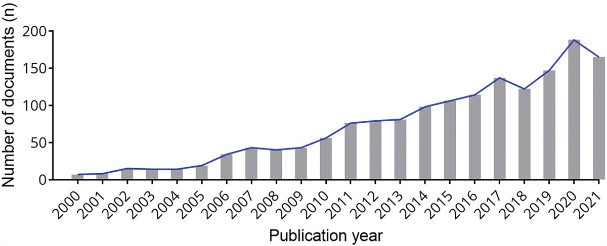
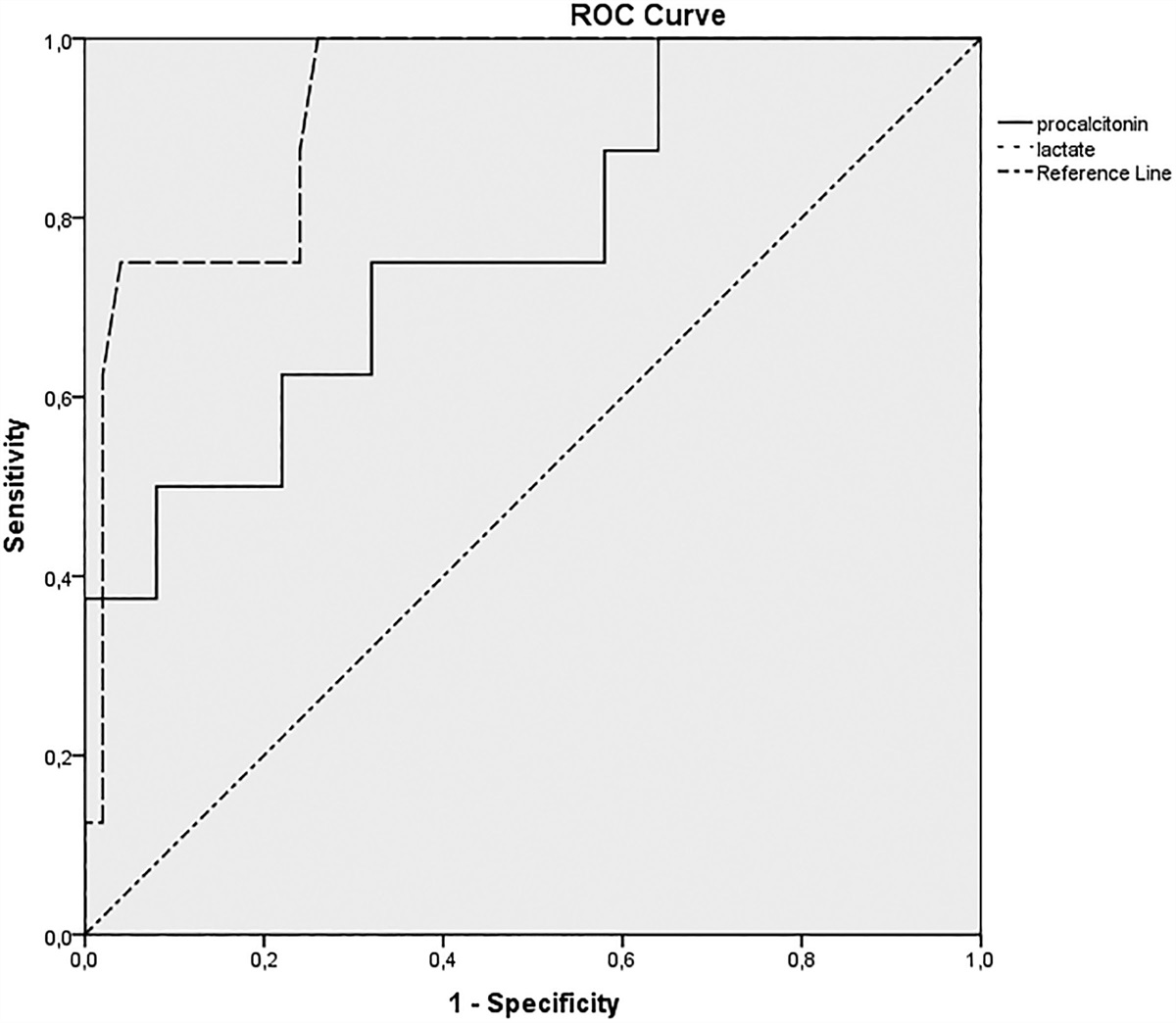
Percentage (%) denotes the prevalence of the risk factors. The data expressed in the table were obtained from original research works and review literature.[5,12,14–36]The International Study on Cerebral Vein and Dural Sinus Thrombosis documented the occurrence of CVT in different venous sinuses: superior sagittal sinus (62%), transverse sinus (41–45%), straight sinus (18%), cortical veins (17.1%), jugular veins (12%), a vein of Galen, and internal cerebral vein (11%) (Fig. 1).[5] Furthermore, in recent studies authors also reported higher incidence of superior sagittal sinus (SSS) involvement 65%,[8] 51%,[37] and 45%[38] cases and transverse sinus was 60.5%,[8] 56%,[37] and 62%[38] patients. Authors also reported multiple venous sinus involvement in 71.2%,[8] and 46%[37] cases. The superficial venous circulation has numerous anastomoses and collateral circulation with variation in the course, which may explain the better prognosis of CVT involving the superficial venous system.[7–12] However, the deep venous system is usually consistent and visible at angiography; thus, thrombosis in the deep venous sinus can be diagnosed easily.[2–6,8–12]
 Figure 1.: Anatomy of Dural venous sinus with distribution of CVT in percentage (Ferro et al[5]). CVT = cerebral venous thrombosis.
We searched electronic databases, especially MEDLINE, EMBASE, CINAHL, and Web of Science collection up to July 2023 to search literature on the epidemiology, clinical features, diagnostic modalities, treatment protocols, and prognosis of cerebral venous thrombosis utilizing medical subject headings terms and Boolean operators to combine search terms. This literature review aims to summarize the current knowledge on the epidemiology, pathophysiology and management of adult CVT.
2. PathophysiologyThe entire pathophysiology has not been experimentally proven, but CVT may present as either of the following 4 distinct clinical syndromes: Intracranial hypertension; Focal neurological syndrome; Diffuse encephalopathy, and; Cavernous sinus syndrome.[2–4,6] The pathophysiological changes in CVT evolve slowly over hours or days and can progress sufficiently for weeks to cause signs and symptoms of CVT. Cerebral vein thrombosis increases venous pressure and reduces capillary perfusion pressure, leading to a rise in cerebral blood volume; ultimately, patients develop intracranial hypertension.[8–12] However, cortical collateral circulation is engaged, but intracranial hypertension subsequently leads to disruption of the blood-brain barrier and the development of vasogenic edema. This pathophysiology causes failure of the sodium-potassium ATPase dependent pump, an indirect regulator of intracellular water volume results in cytotoxic edema development.[3,10–14,37]Figure 2 depicts the pathophysiological changes in CVT.
 Figure 2.: The flowchart illustrates the pathophysiological changes in CVT. The data for constructing this flowchart were obtained from the original studies evaluating the adult CVT population.[2,3,10,11,14,37] CVT = cerebral venous thrombosis.
Superficial cortical veins drain into the SSS against the blood flow within the sinus, resulting in blood turbulence which is further aggravated by the existing fibrous septa at the inferior angle of the sinus. This is the most acceptable explanation of the higher prevalence of thrombosis in SSS.[3–6,14] Furthermore, in addition to draining the cerebral hemisphere, the SSS and other dural venous sinuses also drain blood from diploic, meningeal and emissary veins. This explains the relationship between the occurrence of CVT following infective pathologies in their draining areas, For example, cavernous sinus thrombosis in facial infections, lateral sinus thrombosis in chronic otitis media and sagittal sinus thrombosis in scalp infections.[3,12–14,37]
The dural venous sinuses contain most of the arachnoid villi and granulations, especially in the SSS, responsible for cerebrospinal fluid absorption. So, thrombosis of the dural venous sinus causes blockage of villi and granulations and prevention of cerebrospinal fluid absorption, which eventually leads to intracranial hypertension and papilloedema provoked coma and mortality.[14,37–40]
3. Clinical presentationsThe clinical presentation of CVT is often vague and largely depends on the site and extent of the lesion, age of onset, and associated comorbidities.[8–12] CVT patients may present with a constellation of symptoms, which broadly categorize either as isolated such as intracranial hypertension (ICH) and a focal brain lesion, or a combination of both based on the extent of ICH and brain parenchymal lesion.[15,16,37–40] However, about 40% of patients present with acute stroke-like syndrome within 48 hours of onset, and acute or subacute headache is the most common clinical presentation of CVT, often with a normal neurological finding. The most common clinical presentations are signs of intracranial hypertension and parenchymal drainage: headache (70–90%), seizure (30–40%), papilloedema (30–60%), focal neurological deficits (30–50%), aphasia (15–20%), altered level of consciousness (15–25%), coma (5–15%), and rarely movement disorder.[16–18]
The clinical symptoms corresponding to each type of dural venous sinus thrombosis or overviewed in Table 2. Physicians should be alert for CVT if a patient presents with the following potential symptoms:[16,18,29,42,43]
Table 2 - Clinical presentations according to the affected dural venous sinuses.[2–6,11–14,18,29,30,37–39,41–43] Site of CVT Clinical presentation Superior sagittal sinus (39–62%) Cranial nerve palsies and intracranial hypertension lead to common symptoms:CVT = cerebral venous thrombosis, TS = transverse sinus.
The diagnosis of CVT is based on a high degree of clinical suspicion confirmed by either computed tomography (CT) or magnetic resonance imaging (MRI) with contrast-enhanced venography to demonstrate venous sinus thrombosis.[5,14,37–40] The radiological findings of CVT can be direct visualization of venous sinus without blood flow; or maybe ischemic changes associated with the venous outflow obstruction.[3,11,18] There is no specific laboratory test that can positively exclude CVT in the acute phase of the disease, and blood tests are performed to evaluate coagulation abnormalities like an underlying hypercoagulable state, systemic infection, or an inflammatory process. Furthermore, screening for potential prothrombotic conditions that may predispose to CVT is recommended.[6,8,12,27–30] Details of the radiological findings of adult CVT are illustrated in Table 3.
Table 3 - At a glance merits and demerits of CT, MRI, and DSA techniques.[3,5,11,12,27,44–50] Techniques Traits Description CT Venography Advantages 1.Good visualization of major venous sinusesAV = arteriovenous, CT = computed tomography, CKD = Chronic kidney disease, CVT = Cerebral venous thrombosis, CTV = CT venography, DSA = digital subtraction angiography, MRI = magnetic resonance imaging, MRV = magnetic resonance venography, TOF = time-of flight.
Prompt investigation with an unenhanced CT scan of the brain is the noninvasive imaging method of choice when CVT is clinically suspected. Acute CVT may demonstrate an elongated hyper-attenuating clot known as a “cord sign,” which may persist for 2 weeks and then become isodense to brain parenchyma.[18,40] Generally, a non-contrast CT scan produces an indirect sign that includes the early and late signs of venous ischemia known as sulcal effacement and diffuse parenchymal edema, ventricular effacement, or diminished differentiation between gray and white matter. However, a cerebral infarct not following a typical arterial territory, involving only a subcortical area, multiple unilateral and bilateral lesions with or without hemorrhagic changes should raise a concern about the venous origin.[3,5,12] Further, a cerebral infarct comprising multiple arterial territories should raise concerns about potential venous pathology, particularly CVT.[5,12,27]
CT venography (CTV) is particularly useful in acute and emergency cases and can be utilized as the initial test for assessing the patency of the deep and cortical venous system in a comatose or uncooperative patient.[8–11,18] The most frequent findings on CTV is vascular filling defects and an “empty delta sign” when the superior sagittal sinus is involved.[44,45] However, an artifact from dense cortical bones significantly reduces the diagnostic accuracy of the CT venography, and also, arachnoid granulations may protrude into the venous sinuses, mimicking filling defects by thrombus, which is another potential disadvantage of CTV imaging.[44–47] In infants, a false dense clot sign may result from the relatively high density of the blood in the sagittal sinus, and a false, empty delta sign may cause hyperdense empyema.[5–10,48] Occasionally, engorged and dilated venous malformations produce a hyperdense lesion on unenhanced CT and demonstrate a characteristic linear enhancing focus converging on a single dilated vein known as “caput medusa” or “candelabra” appearance on CT venography.[3,5–12,18,44]
4.3. MRI and MR venographyConventional T1 and T2 weighted MRI is more sensitive than an unenhanced CT scan to diagnose a case of CVT.[45–47] On standard sequences, the early signs include the absence of a typical venous flow pattern and abnormal signal within the dural venous sinus. A brief description of the evolution of thrombus signal intensity caused by the paramagnetic effects of hemoglobin degradation products is provided in Table 3.
Magnetic resonance venography (MRV) is helpful in either acute or subacute and emergency or ambulatory cases and to confirm suspected cases of deep venous thrombosis where CT venography was inconclusive or normal.[5,7–12,40,44,45] Contrast-enhanced MRV offers improved visualization of the cerebral venous system and is unlikely to be affected by complex blood flow.[8,18,42] However, in MRI venography, aplasia and hypoplasia of the transverse sinus can be mistaken. There is also the chance of signal loss due to in-plane flow, and hyperintense thrombi can mimic patent sinus during time-of-flight angiography. Nonetheless, both MR venography and CT venography are adequate for CVT diagnosis, but MRV has higher diagnostic accuracy for the visualization of brain parenchymal lesions.[6,41–45]
4.4. Digital subtraction angiography (DSA)Although DSA is considered a gold standard technique it usually only performed in the presence of either unclear CTV and MRV imaging or when endovascular intervention is planned because of its associated risk.[29,44–49] Generally, there are filling defects in the dural venous sinuses or cortical veins, delayed venous drainage, and dilated collateral circulation. There may also be an abrupt cutoff of cortical veins with surrounding tortuous and dilated “corkscrew” collateral circulation.[5,18,29] Furthermore, DSA can identify vascular aneurysm and dural arteriovenous fistula, which might cause the formation of a false “corkscrew” sign due to sluggish venous drainage and vascular congestion.[50] Nevertheless, DSA has a unique ability to measure venous pressure and pressure > 10 mm H2O indicates a probability of parenchymal damage, which carries significant value for treatment outcome.[41,45]
5. Treatment and guidelines 5.1. Overview of treatment protocolsPrompt diagnosis to identify and treat the associated factors, initiate anticoagulation therapy, and manage ICH should maximize the chance of a favorable outcome.[18,39,41] The management algorithm of CVT is illustrated in Figure 3.
Comments (0)