Study design and population
The Review Board of Peking University People’s Hospital approved the study protocol (No. 2021PHB237-001). We prospectively registered the study prior to patient enrolment at the Chinese Clinical Trials Registry (ChiCTR2100052810)Footnote 1 on 5 November 2021. This study was performed in accordance with the Helsinki Declaration and the manuscript follows the guidelines of Consolidated Standards of Reporting Trials (CONSORT). Written informed consent was obtained from patients before participation in the study. The patients were allowed to withdraw their consent and cease participation in the study at any time.
The inclusion criteria were patients from 18 to 75 yr of either sex who were scheduled for MIDCAB. The exclusion criteria were a New York Heart Association functional classification of IV, a preoperative ejection fraction of < 40%, a body mass index of < 18 or > 35 kg·m−2, redo or emergency cases, coagulopathy, localized infection at the block site, allergy or intolerance to local anesthetics, hepatic or renal insufficiency, diagnosed mental disorder, history of chronic pain conditions requiring regular opioids, and refusal to participate.
After provision of written informed consent, 60 participants were enrolled in the study between November 2021 and June 2022.
Randomization and blinding
Randomization was conducted in a 1:1 ratio according to computer-generated randomized numbers. A nurse anesthetist who had no involvement in the collection of perioperative data or statistical analysis performed the randomization and sealed the group assignments in sequentially numbered opaque envelopes. The envelope was opened by the nurse anesthetist one hour before surgery and then a 30-mL syringe of the study medication was prepared according to the allocation number in the envelope. The patients randomly allocated to the ESPB group received 0.5% ropivacaine, whereas the control group received 0.9% normal saline. Both syringes were identical and labelled as “study medication” to ensure blinding. All patients, anesthesiologists who performed the block and provided patient care, nursing staff, cardiac surgeons, and investigators were unaware of the group allocations.
Interventions
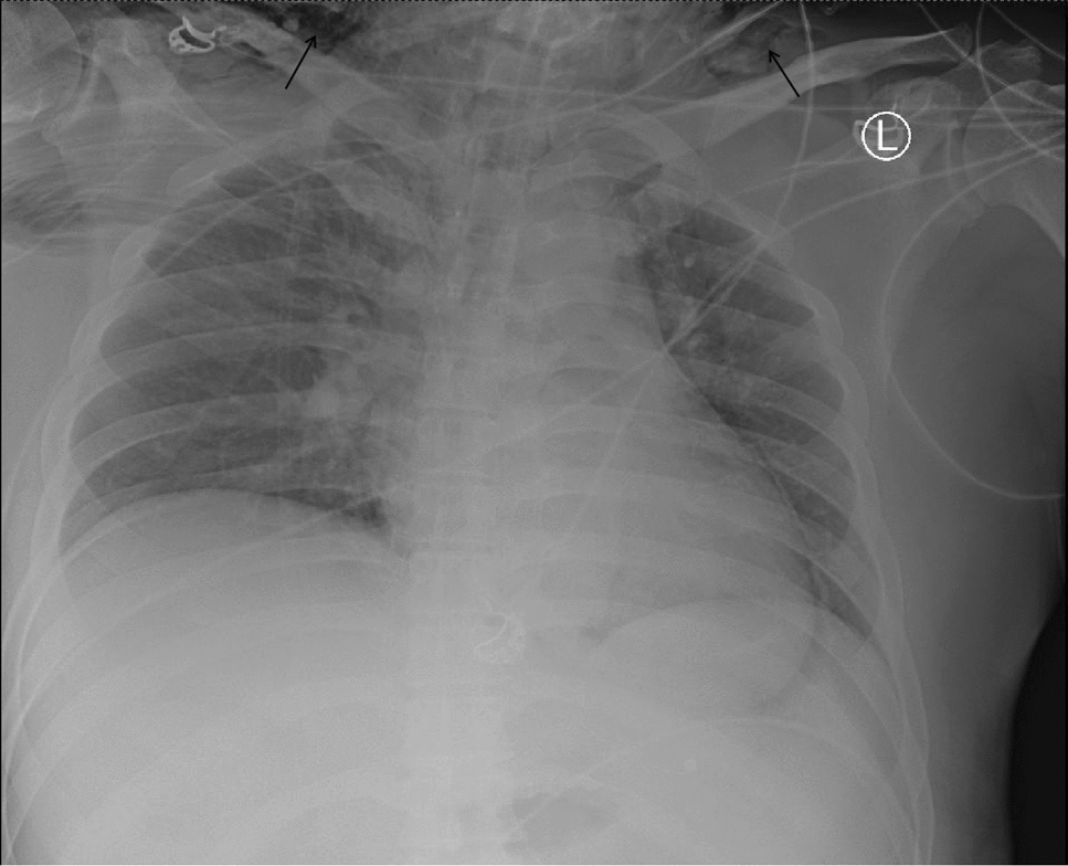
All the ESPBs were performed by the same experienced attending anesthesiologist before general anesthesia induction. The patient was set in the right lateral position under sedation by iv midazolam (1–2 mg). After aseptic preparation of the skin area with chlorhexidine gluconate, a low-frequency linear transducer (Logiq™ e, GE HealthCare, Chicago, IL, USA) in a sterile cover was placed in the paramedian sagittal plane at the T5 level, approximately 2–3 cm lateral to the posterior midline. Then, the probe was adjusted to visualize the transverse process with acoustic shadow and the erector spinae muscle above it. A block needle (22G, 100 mm, Plexufix®, B. Braun SE, Melsungen, Germany) was then introduced from the cephalad to caudad under local anesthetic infiltration using the in-plane technique. The needle target was the interfascial space between the transverse process and erector spinae muscle. The correct placement of the needle tip was verified by 1–2 mL 0.9% saline, which was confirmed as interfascial spread of injectate lifting the erector spinae muscle. Then, 30 mL of 0.5% ropivacaine or 0.9% normal saline was injected.
Perioperative management
Patients were monitored with 5-lead electrocardiogram, pulse oxygen saturation, and invasive blood pressure monitoring. General anesthesia induction included a combination of iv midazolam (0.03–0.05 mg·kg−1), etomidate (0.15–0.2 mg·kg−1), cis-atracurium (0.15–0.2 mg·kg−1), and fentanyl (3–5 µg·kg−1). Then, a left-side double-lumen tube with appropriate size was inserted. After induction, a 7-Fr central venous catheter with triple-lumen was placed under ultrasound guidance. Propofol (3–5 mg·kg−1·hr−1), cisatracurium (0.1–0.2 mg·kg−1·hr−1), and dexmedetomidine (0.5–0.8 µg·kg−1·hr−1) were used for anesthesia maintenance to achieve a bispectral index between 40 and 60. In case of hypertension (defined as a blood pressure > 20% of baseline value), the anesthesia depth of the patient was checked, then propofol infusion rate was adjusted and/or fentanyl boluses (1–2 µg·kg−1) were administered by the attending anesthesiologist. A phenylephrine or dopamine bolus and/or infusion was used if the blood pressure was < 20% of the baseline value though decreasing propofol infusion rate or fluid volume loading was applied. No other opioids except fentanyl were used during surgery. The anesthesiologists who provided intraoperative care were not otherwise involved in the data collection for this study.
All iv anesthetic infusions were stopped once surgery was completed, and the patient was transferred to the cardiac ICU. The medical staff in the ICU performed extubation once the patient fulfilled weaning criteria. No analgesic infusions were administered in the cardiac ICU until the patient was extubated.
Patient-controlled analgesia (PCA) with iv hydromorphone was prescribed with no basal infusion and an intermittent bolus dose of 0.2 mg and a lock-out period of ten minutes. Intravenous tropisetron 5 mg was given to all patients after completion of surgery. After being transferred to the ward, further analgesic treatment with oxycodone and acetaminophen was prescribed if necessary.
Surgical procedure
The same cardiac surgical team performed all the procedures. Thoracotomy was performed with the patient positioned supine and the left shoulder elevated at 30°, then a 5–6-cm curvilinear incision was performed between the fourth and fifth ribs on the left anterolateral chest wall. The internal mammary artery was obtained using a retractor system via direct vision. With the employment of Octopus® Nuvo Tissue Stabilizer (Medtronic, Dublin, Ireland) and Starfish™ NS Heart Positioner (Medtronic, Dublin, Ireland) to achieve stabilization, the anastomosis between the distal graft and the target vessel was performed on the beating heart.15
Outcomes
Our primary outcome was the numerical rating scale (NRS) pain scores at rest within 48 hr postoperatively (i.e., after skin closure; assessed at six hours, 12 hr [used for sample size calculation; cf. Statistical analysis below], 18 hr, 24 hr, and 48 hr). The secondary outcomes were NRS pain scores on deep inspiration at the same time points after skin closure, postoperative hydromorphone consumption, and quality of recovery-15 (QoR-15) scores at 24 hr and 48 hr after surgery. Other outcomes included total fentanyl use, time to tracheal extubation and chest tube removal after surgery, ICU length of stay, and time to hospital discharge. Adverse events including postoperative nausea and vomiting (PONV), atrial fibrillation, pleural effusion, pericarditis, and block-related complications such as hematoma, pneumothorax, infection, and 30-day mortality were recorded.
The magnitude of postoperative pain was evaluated at six hours, 12 hr, 18 hr, 24 hr, and 48 hr by a follow-up investigator who was blinded to the study group allocations using the NRS score (0 = no pain, while 10 = worst pain ever). Analgesic consumption and the QoR-15 score were collected at 24 hr and 48 hr after surgery. We used the Chinese version of the QoR-15 questionnaire as a patient-centred measurement of recovery quality. This questionnaire contains the following domains: physical comfort and independence, pain, psychological state, and emotional state.16 A QoR-15 score of 0 represents poor recovery while a score of 150 represents excellent recovery. Adverse events, postoperative complications, and patient progression were traced by visiting the patients daily or reviewing electronic medical records during their entire hospital stay. A telephone interview was performed at one month after surgery to assess 30-day mortality.
Statistical analysis
The sample size was calculated with PASS power analysis and sample size software (NCSS LLC., Kaysville, UT, USA). Based on results from a pilot study of ten patients, the mean and standard deviation (SD) of postoperative NRS scores at 12 hr in patients using iv hydromorphone PCA were 3.7 and 1.9, respectively. Thus, to identify a relevant clinical difference of 1.5 in the NRS score with a two-sided 5% significance level, 23 patients were calculated in each group to have 90% power using a repeated measures analysis of variance (ANOVA) method with five observations on the same subject. Autocorrelation between repeated observations on each subject was assumed to be 0.6. Considering possible dropouts, we increased the sample size to 30 patients for each study group.
Statistical analysis was performed using IBM SPSS Statistics for Windows version 24.0 (IBM Corp., Armonk, NY, USA). Before analysis, the Shapiro–Wilk test was employed to analyze the normality of data distribution. Continuous data are presented as mean (SD) or median [interquartile range (IQR)]. Dichotomous and polytomous data are presented as counts and percentages. For NRS scores, between-group differences at all time points after surgery were analyzed using two-way repeated measures ANOVA with Bonferroni correction. Confidence intervals (CIs) and P values are reported after Bonferroni adjustment for the 5-time comparisons, so are presented as 99% CIs. For the other outcomes, the Mann–Whitney U test or independent samples t test was adopted to compare continuous variables. Categorical data were analyzed by the Chi square or Fisher's exact test when necessary. All P values were two sided and an adjusted P value less than 0.05 was considered statistically significant for the primary outcomes. For secondary outcomes, a P < 0.05 was considered significant.


Comments (0)