
Remember me

N-Acylhydrazones are a class of compounds that contain the hydrazonic functional group (–NH–N=C–) attached to an acyl group, which can be modified to generate a range of different structures with varying properties . The versatility of this class of compounds is also related to the ability of N-acylhydrazones to exist as different isomers and/or tautomers. They can exist as geometric isomers (E/Z), which differ in the orientation of the groups around the carbon–nitrogen double bond , as well as amido and iminol tautomers . The ability to undergo E/Z isomerization in a stimuli-responsive imine bond is what makes this class useful for applications in the field of molecular electronics, as switchers . In addition, these compounds can also adopt syn- or antiperiplanar conformations, due to the constriction of the rotation around the conjugated amide single bond (N–C=O).
N-Acylhydrazones have also gained attention in literature due to other applications, ranging from medicine to supramolecular chemistry . Among their applicability is the area of optoelectronic devices, in which they are used for the manufacture of organic light-emitting diodes (OLEDs) . Moreover, studies involving N-acylhydrazonic derivatives have highlighted their suitability for the treatment of pathologies associated with infection and/or inflammation . Antimicrobial activity is one of the most frequently studied and reported biological properties of this class . Angelova and co-workers, for example, reported the ability of sulfonyl hydrazones and 4-methyl-1,2,3-thiadiazole-based hydrazone derivatives to inhibit the growth of several bacterial strains by interfering with their metabolism or cell membrane integrity .
In the context of cancer therapy development, metal complexes of N-acylhydrazones stand out. For example, Firmino et al. demonstrated that gallium(III) complexes of isoniazid-derived hydrazones exhibit strong cytotoxicity against HL-60 and HCT-116 cancer cell lines . The study also found that those coordination compounds were selective towards abnormal cells, exhibiting lower toxicity for healthy human hepatocytes. On the other hand, an important development in cancer research is the use of physiological metal ion complexes, which afford more biocompatibility and thus less side-effects in therapy . In this sense, we have reported dicopper(II) complexes from different N-acylhydrazonic binucleating ligands with potent antiproliferative activity against a panel of cancer cell lines .
On the field of neurodegeneration, our research group was the first to establish the suitability of N-acylhydrazones as novel metallophores able to affect protein aggregation and/or oxidation enhanced by physiological metal–protein anomalous interactions related to Alzheimer's (AD) and Parkinson's (PD), as well as to prion diseases . Our lead compound INHHQ (or 8-hydroxyquinoline-2-carboxaldehyde isonicotinoyl hydrazone) has been successfully tested in the prevention of short- and long-term memory deficits in a mice model of sporadic AD . Additionally, INHHQ decreases copper-mediated production of reactive oxygen species (ROS) in vitro, which may be another mechanism through which the compound exerts its protective effects in the brain.
From a drug development perspective, however, INHHQ has some pharmacological limitations, such as low solubility and certain susceptibility to hydrolysis in a water-rich medium. The protective character and high metallophoric potential of INHHQ, nevertheless, prompted us to design and synthesize new optimized derivatives. In this context, we recently described a family of pharmocologically improved N-acylhydrazones containing the 1-methylimidazole moiety . In 2020, we proved the promising anti-PD and metallophoric effect, especially towards intracellularly relevant copper(I) ions, of X1INH (1-methyl-1H-imidazole-2-carboxaldehyde isonicotinoyl hydrazone) .
This year, we evaluated the effects of the presence of three methoxy substituents in an N-acylhydrazone derived from 3,4,5-trimethoxybenzoic acid hydrazide, a modification inspired by mescaline, the active principle of the hallucinogenic cactus peyote, which could result in a greater BBB penetration . In this study, however, the structural modifications in the compound did not seem to significantly affect its pharmacological properties and metallophoric potential against copper(II) when compared to the unsubstituted counterpart. Nevertheless, the bioinspired compound was still able to reduce oxidative stress and affect the aggregation of the amyloid-β peptide, related to pathophysiological events in AD.
As a continuation of our long-term effort on the development of potentially bioactive N-acylhydrazones, the present work comprises a structural and spectroscopic comparison, from both experimental and theoretical viewpoints, of two structure-related hydrazones. Both compounds are new, and derived from the same 3,4,5-trimethoxybenzoic acid hydrazide, but differ with respect to the carbonyl precursors: herein, 3-substituted salicylaldehydes (Scheme 1) are used, which assure for a harder donor-atoms set in order to target trivalent metal ions such as aluminum(III), which has been proposed to display a role in neurodegeneration .
![[1860-5397-19-125-i1]](https://www.beilstein-journals.org/bjoc/content/inline/1860-5397-19-125-i1.svg?scale=2.0&max-width=1024&background=FFFFFF)
Scheme 1: Structure of (A) 3-methylsalicylaldehyde 3,4,5-trimethoxybenzoyl hydrazone (hdz-CH3) and (B) 3-nitrosalicylaldehyde 3,4,5-trimethoxybenzoyl hydrazone (hdz-NO2).
A comparative study between these two N-acylhydrazones is interesting, especially considering that they possess different substituents at the same position in the phenol ring: the electron-donating methyl group (hdz-CH3) and the electron-withdrawing nitro group (hdz-NO2). It is expected that those substituents impact the chemical (e.g., acidity and hydrolysis susceptibility) as well as the structural and spectroscopic properties of the compounds.
Results and DiscussionThe methyl-substituted hdz-CH3 and its nitro-containing analogue hdz-NO2 were isolated as beige and light-yellow solids with 78% and 44% yield, respectively. Thermal analyses between 25 and 350 °C were performed in order to verify the hydration status of the bulk. Regarding hdz-CH3, a weight loss of 9.78% from around 80 to 190 °C was observed, suggesting the presence of two crystallization water molecules in the network (calcd.: 9.47% for C18H20O5N2·2H2O, MW = 380.39 g mol−1). On the other hand, hdz-NO2 did not show any mass loss below 250 °C, indicating the absence of solvation molecules in the sample (C17H17O7N3, MW = 375.34 g mol−1).
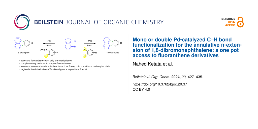
Single crystals of both compounds, as monohydrates, were isolated from the respective mother liquors. The structures of hdz-CH3 and hdz-NO2 are displayed in Figure 1 and an overview of the crystallographic data can be found in Table 1.
![[1860-5397-19-125-1]](https://www.beilstein-journals.org/bjoc/content/figures/1860-5397-19-125-1.png?scale=2.0&max-width=1024&background=FFFFFF)
Figure 1: ORTEP representation of the new N-acylhydrazones synthesized in the present work, drawn with thermal ellipsoids at 30% probability. Left: hdz-CH3 and right: hdz-NO2.
Table 1: Crystal, data collection and refinement parameters for hdz-CH3 and hdz-NO2.a
Data hdz-CH3 hdz-NO2 crystal size (mm) 0.99 × 0.14 × 0.14 0.57 × 0.32 × 0.12 empirical formula C18H22N2O6 C17H19N3O8 formula weight (g mol−1) 362.37 393.35 F(000) 1536 412 temperature (K) 293 293 absorption coefficient μ (mm−1) 0.101 0.117 calculated density (g·cm−3) 1.337 1.452 crystal system monoclinic triclinic space group C2/c (n° 15)![[Graphic 1]](https://www.beilstein-journals.org/bjoc/content/inline/1860-5397-19-125-i3.svg?max-width=637&scale=1.0) (n° 2)
a, b, c (Å)
32.1401(15)
(n° 2)
a, b, c (Å)
32.1401(15)
aa, b, c, α, β, γ: unit cell parameters; Z: formula unit per unit cell; Z’: number of formula units in the crystallographic unit cell divided by the number of independent general positions; F(000): structure factor in the zeroth-order case; F: structure factor; F2: squared structure factor; T: transmission factor.
Both molecules are near planar and correspond to the (E)-isomer, in an antiperiplanar conformation. Superposition of the structures (Figure 2A) shows that spatial arrangements are nearly the same and even the crystallization water molecules were allocated in nearby sites, interacting as H-acceptors in a hydrogen bond with the respective N2H groups. In spite of these similarities, the compounds were indexed in different space groups. The methyl-containing hdz-CH3 crystallized in the monoclinic system, C2/c space group, while the nitro derivative hdz-NO2 belongs to the ![[Graphic 2]](https://www.beilstein-journals.org/bjoc/content/inline/1860-5397-19-125-i4.svg?max-width=637&scale=1.18182) group, from the triclinic system. In both cases, a moderate to strong intramolecular H-bond involving the phenol oxygen O1 as H-donor and the azomethine nitrogen N1 as H-acceptor is observed, which originates six-membered cyclic motifs with a graph-set
group, from the triclinic system. In both cases, a moderate to strong intramolecular H-bond involving the phenol oxygen O1 as H-donor and the azomethine nitrogen N1 as H-acceptor is observed, which originates six-membered cyclic motifs with a graph-set ![[Graphic 3]](https://www.beilstein-journals.org/bjoc/content/inline/1860-5397-19-125-i5.svg?max-width=637&scale=1.18182) (6) . The hydrogen atom is closer to O1 than to N1, indicating that the preferred protonation site is the former, as can be observed at the Fourier difference maps (Figure S1A and S1B in Supporting Information File 1), but the presence of the hydrogen bond influences the contiguous aromatic system, as can be inferred by the characteristic elongation of the C1–C2 bond, which is 1.405(2) Å in hdz-CH3 and 1.411(3) Å in hdz-NO2 . The effect is also noticed through the HOMA (harmonic oscillator model of aromaticity) indexes of the rings: while the trimethoxy-substituted, hydrazide-derived one presents values of 0.995 (hdz-CH3) and 0.990 (hdz-NO2), indicating high aromaticity, the aldehyde-derived ring shows HOMA indexes of 0.964 (hdz-CH3) and 0.961 (hdz-NO2), suggesting that, for both compounds, this H-bond decreases the aromaticity of the phenol-containing ring. Finally, O1···N1 distances are 2.581(2) and 2.539(3) Å for hdz-CH3 and hdz-NO2, respectively (Table 2). Thus, all the structural evidence discussed above converges to the conclusion that the presence of the electron-withdrawing nitro substituent in hdz-NO2 makes the intramolecular H-bond stronger.
(6) . The hydrogen atom is closer to O1 than to N1, indicating that the preferred protonation site is the former, as can be observed at the Fourier difference maps (Figure S1A and S1B in Supporting Information File 1), but the presence of the hydrogen bond influences the contiguous aromatic system, as can be inferred by the characteristic elongation of the C1–C2 bond, which is 1.405(2) Å in hdz-CH3 and 1.411(3) Å in hdz-NO2 . The effect is also noticed through the HOMA (harmonic oscillator model of aromaticity) indexes of the rings: while the trimethoxy-substituted, hydrazide-derived one presents values of 0.995 (hdz-CH3) and 0.990 (hdz-NO2), indicating high aromaticity, the aldehyde-derived ring shows HOMA indexes of 0.964 (hdz-CH3) and 0.961 (hdz-NO2), suggesting that, for both compounds, this H-bond decreases the aromaticity of the phenol-containing ring. Finally, O1···N1 distances are 2.581(2) and 2.539(3) Å for hdz-CH3 and hdz-NO2, respectively (Table 2). Thus, all the structural evidence discussed above converges to the conclusion that the presence of the electron-withdrawing nitro substituent in hdz-NO2 makes the intramolecular H-bond stronger.
![[1860-5397-19-125-2]](https://www.beilstein-journals.org/bjoc/content/figures/1860-5397-19-125-2.png?scale=2.0&max-width=1024&background=FFFFFF)
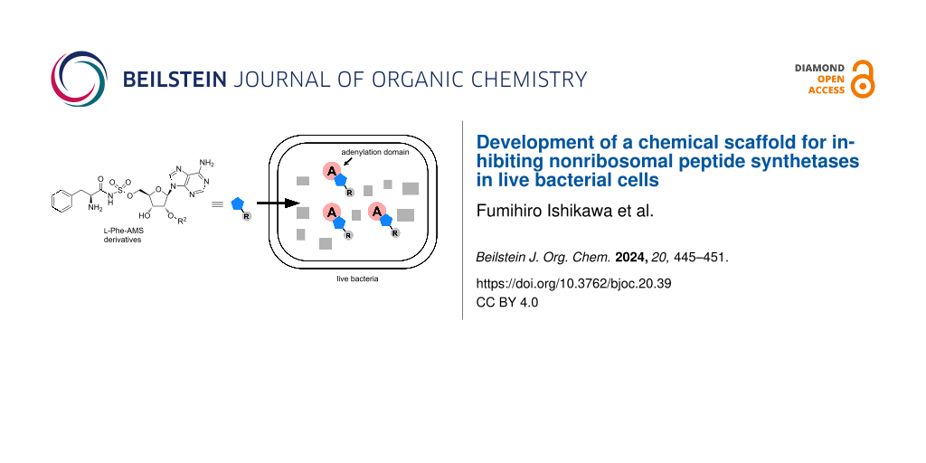
Figure 2: (A) Superposition of molecular structures and stacked motifs of (B) hdz-CH3 and (C) hdz-NO2.
Table 2: H-bonding parameters for hdz-CH3 and hdz-NO2.
D–H···A Symmetry operation D–H (Å) H···A (Å) D···A (Å) D–H···A (°) % ΣvdWr hdz-CH3 O1–H1···N1 intra 0.93(2) 1.772(18) 2.581(2) 144(2) 64.4 Ow–Hw1···O2 – 0.84(3) 2.00(3) 2.816(2) 166(3) 73.5 N2–H2a···Ow 1−x,1−y,1−z 0.840(17) 2.164(18) 2.910(2) 148(2) 79.6 C14–H14···O2 intra 0.9300 2.400 2.734(2) 101.00 87.3 hdz-NO2a O1–H1···N1 intra 0.86(4) 1.79(3) 2.539(3) 146(3) 65.1 Ow–Hw2···O4 – 0.83(5) 2.03(5) 2.839(4) 169(5) 74.6 Ow–Hw1···O2A 1−x , −y, 1−z 0.82(4) 2.48(5) 3.143(13) 138(4) 91.2 Ow–Hw1···O1 1−x , −y , 1−z 0.82(4) 2.34(4) 3.061(3) 147(4) 86.0 N2–H2a···Ow 1−x , 1−y , 1−z 0.83(2) 2.09(2) 2.898(4) 163(3) 76.8 C5–H5···O2 −1+x , y , 1+z 0.9300 2.5800 3.175(4) 162.00 94.8 C10–H10···Ow 1−x , 1−y , 1−z 0.9300 2.4600 3.360(4) 162.00 91.2 C14–H14···O4 intra 0.9300 2.4400 7.758(4) 100.00 89.7 C16–H16c···O5 intra 0.9600 2.4100 2.975(5) 117.00 88.6 C17–H17b···O3 1−x , −y , 1−z 0.9600 2.5900 3.536(5) 168.00 95.2aData shown only for the major component of disorder.
Regarding lattice organization, both structures exhibit stacked motifs. In hdz-CH3, the ![[Graphic 4]](https://www.beilstein-journals.org/bjoc/content/inline/1860-5397-19-125-i6.svg?max-width=637&scale=1.18182) planes were organized by C–H···O non-conventional H-bonds comprising the methoxy groups (Figure 2B). In the columns, there are antiparallel dimers (marked as *) sandwiched between two non-parallel molecules. The planes in hdz-NO2 present similar intermolecular interactions, but they grow as (101) planes. The columns also present the antiparallel dimers, but they are intercalated by antiparallel slipped molecules (Figure 2C).
planes were organized by C–H···O non-conventional H-bonds comprising the methoxy groups (Figure 2B). In the columns, there are antiparallel dimers (marked as *) sandwiched between two non-parallel molecules. The planes in hdz-NO2 present similar intermolecular interactions, but they grow as (101) planes. The columns also present the antiparallel dimers, but they are intercalated by antiparallel slipped molecules (Figure 2C).
The supramolecular organization of the N-acylhydrazones was also studied through their Hirshfeld surfaces, in which π–π interactions play an important role. When the normalized distance between a molecule and the closest neighbors are plotted (Figures S2A and S2C, and S3A and S3C in Supporting Information File 1), it becomes possible to calculate the contribution of each type of intermolecular interaction to the whole profile . In our case, results show that the sum of π···π and C–H···π contacts comprises almost 20% of the Hirshfeld surface for both compounds. The curvedness maps were also plotted over the Hirshfeld surfaces and evidence the strong flatness of the structures (Figures S2B and S3B in Supporting Information File 1). Important contributions of hydrogen bonds (O···H/H···O) can also be found, especially for hdz-NO2. Besides the interaction maps, crystal structures themselves may be used to calculate the electrostatic potential over an electronic density map as well. For hdz-NO2, results show the alternated slipped stack motif involves the assembly of portions with opposite electrostatic potentials.
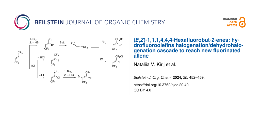
Computational calculations were performed in order to confirm some of the structural and vibrational aspects of the compounds. The optimized structures obtained with B3LYP/6-311G(d,p) (both in gas and water phases) were compared to the respective experimental XRD structures (Figure 3). The RMSD values for hdz-NO2 in gas (0.431 Å) and water (0.405 Å) phases were lower than the respective values for hdz-CH3 (0.648 Å in the gas phase and 0.623 Å in water). However, the small RMSD values in all cases showed that the calculated structures correlated pretty well with the respective experimental ones . The biggest differences between the theoretical structures and the experimental data corresponded to the aldehyde-derived ring, containing the –CH3 (or –NO2) substituents, an effect which is more explicit in hdz-NO2, probably due to the intrinsic disorder observed in its structure.
![[1860-5397-19-125-3]](https://www.beilstein-journals.org/bjoc/content/figures/1860-5397-19-125-3.png?scale=2.0&max-width=1024&background=FFFFFF)
Figure 3: Overlap of the experimental (carbon atoms colored in gray) and theoretical structures (calculated with B3LYP/6-311G(d,p) in gas and water phases colored in yellow and cyan, respectively): hdz-CH3 seen from (A) above and (B) from the side; hdz-NO2 seen from (C) above and (D) from the side. The optimized structures of hdz-CH3 and hdz-NO2 can be seen in Figure S4 in Supporting Information File 1.
The comparison between theoretical and experimental selected geometric parameters can be seen in Tables S1 and S2 (Supporting Information File 1) and confirms they are in good accordance. For hdz-CH3, a maximum percentage error of 3% for bonds and 2% for angles was observed, while hdz-NO2 presented values of 2% and 3% for these parameters, respectively. In turn, the intramolecular interaction OH···N1 showed maximum errors of 2% (hdz-CH3) and 0% (hdz-NO2). In general, the structures calculated involving the IEFPCM formalism (water phase) displayed the best agreement with the experimental data.
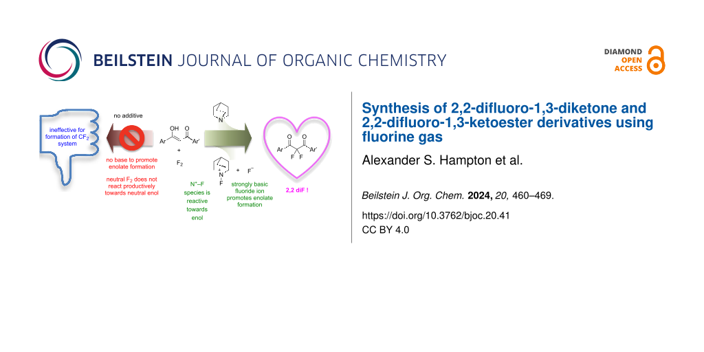
Regarding the vibrational characterization, infrared spectra were also calculated with both B3LYP/6-311G(d,p) – gas phase – and B3LYP/6-311G(d,p)/IEFPCM – water – for hdz-CH3 and hdz-NO2 and fitted very well with the respective experimental data, especially in the lower-frequencies region, i.e., below 2000 cm−1 (Figure 4 and Tables S3 and S4 in Supporting Information File 1). It is worth noting that the assignments of experimental absorptions were performed not only with the aid of DFT calculations, but also checked by comparing them to the vibrations of the respective carbonyl and hydrazide precursors.
![[1860-5397-19-125-4]](https://www.beilstein-journals.org/bjoc/content/figures/1860-5397-19-125-4.png?scale=2.0&max-width=1024&background=FFFFFF)
Figure 4: Mid-infrared spectra of the compounds. Experimental conditions: KBr pellets, room temperature. Calculated conditions: gas phase, level of theory B3LYP/6-311G(d,p). (A) Overlapping of the theoretical (black) and experimental (purple) spectra of hdz-CH3. (B) Overlapping of the theoretical (black) and experimental (yellow) spectra of hdz-NO2.
Although the phenol-related ν(O–H) bands could not be accurately identified due to overlapping with the water stretching modes, theoretical results indicate that the frequency in hdz-CH3 (3407 cm−1) is higher than that in hdz-NO2 (3326 cm−1), suggesting a lower bond force constant in the latter. Differences were observed in the hydroxy bending vibrations as well, and those were perfectly observable in the spectra: δip(C–O–H) and δoop(O–H) modes were assigned, respectively, at 1376 and 720 cm−1 for hdz-CH3, and at 1359 and 747 cm−1 for hdz-NO2. Interestingly, DFT showed that, while these vibrations are “clean” in hdz-CH3, they were coupled with NBA ring movements in hdz-NO2. Therefore, the IR results confirm the stronger character of the intramolecular H-bond in the nitro-substituted N-acylhydrazone. As previously observed for other compounds of this class, the azomethine ν(C=N) modes are barely susceptible to interactions involving the nitrogen atom, being located at 1620 cm−1 in hdz-CH3 and at 1622 cm−1 in compound hdz-NO2.
Other typical bands of N-acylhydrazones were also attributed, such as ν(C=O) at 1658 (hdz-CH3) and 1648 cm−1 (hdz-NO2), as well as ν(N–N) at 1003 (hdz-CH3) and 997 cm−1 (hdz-NO2). The νas(NO2) and νsym(NO2) vibrations of the nitro group in hdz-NO2 were identified as medium and strong intensity bands, respectively, at 1519 and 1336 cm−1. These modes were calculated at 1608 and 1379 cm−1 in the gas phase.
Although N-acylhydrazones are usually prone to undergo speciation in DMSO-d6 solution , 1H NMR measurements showed the existence of only one set of signals in the spectra of hdz-CH3 and hdz-NO2 (Figure 5A and 5B, respectively). 13C NMR and 2D homonuclear (COSY) and heteronuclear (13C,1H-HSQC and HMBC) experiments were employed for the full characterization of these hydrazones, and the spectra can be seen in Supporting Information File 1, Figures S5–S12.
![[1860-5397-19-125-5]](https://www.beilstein-journals.org/bjoc/content/figures/1860-5397-19-125-5.svg?scale=2.0&max-width=1024&background=FFFFFF)
Figure 5: 1H NMR (400 MHz) spectra of (A) hdz-CH3 and (B) hdz-NO2 in DMSO-d6 at 25 °C.
Both compounds exhibit only one NH signal around 12 ppm, indicating the presence exclusively of the (E)-isomer in solution. Furthermore, steric hindrance in these cases allow only for the formation of the antiperiplanar conformation around the conjugated amide single bond. Full assignments, with the chemical shifts and coupling constants, can be found in Table S5 of Supporting Information File 1. The azomethine hydrogen H7 appears as a singlet at 8.58 ppm for hdz-CH3 and is slightly deshielded in the nitro-derivative, resonating at 8.74 ppm, since the electron density-withdrawing substituent in hdz-NO2 increases the electrophilic character of C7.
In both hdz-CH3 and hdz-NO2, methoxy hydrogens occur as a pair of singlets: H15 and H17 at 3.88 ppm, and H16 at 3.74 ppm. Methyl H18 in hdz-CH3 appears as a more shielded singlet at 2.22 ppm. Regarding the aromatic region of this compound, the doublet related to H6 (7.28 ppm) is partially overlapped with the singlet appointed to H10 and H14 (7.27 ppm). These signals were unequivocally assigned using the 2D COSY and HMBC experiments. The most shielded doublet at 7.22 ppm corresponds to H4, and H5 occurs as a triplet at 6.86 ppm. With respect to the aromatic hydrogen atoms of hdz-NO2, the singlet at 7.28 ppm was assigned to the equivalent H10 and H14. In this case, not only there was no overlapping of signals but also H4 appears more deshielded (8.01 ppm) when compared to hdz-CH3. H5, in turn, appears as a triplet at 7.14 ppm.
The presence of the electron-withdrawing NO2-substituent in the aldehyde-derived portion of hdz-NO2 caused a strong deshielding of the hydroxy group (assigned at 12.84 ppm) due to the removal of electron density on the carbon adjacent to –OH. On the other hand, in hdz-CH3, the presence of the methyl substituent moderately shields this proton (11.89 ppm). A comparison of the H7 and –OH chemical shifts of the related hydrazones hdz-CH3 and hdz-NO2 indicates that, also in solution, the intramolecular H-bond is stronger in the latter.
Since N-acylhydrazones may be susceptible to hydrolysis, especially those containing a hydroxy group in ortho-position relative to the azomethine group as the intramolecular H-bond between the phenolic hydrogen and double-bonded nitrogen activates the azomethine carbon for a nucleophilic attack by a solvent molecule, an important step in the development of a new bioactive hydrazonic derivative is the assessment of its stability in aqueous medium. Thus, the electronic absorption spectra of hdz-CH3 and hdz-NO2 were recorded in a 10% DMSO/buffer solution (pH 7.4) immediately after preparation and at regular time intervals.
The UV–vis spectrum of hdz-CH3 between 250 and 450 nm (Figure 6A) shows two multicomponent absorptions centered at 298 (εapp = 20,700 ± 40 L mol−1 cm−1) and 338 nm (10,000 ± 25 L mol−1 cm−1), which could be fitted to the sum of five gaussian bands. Of these, the one at 276 nm and that of very low intensity at 307 nm were tentatively assigned by comparison with the spectra of the precursors to transitions mainly localized in the TMP ring. Nevertheless, a contribution of the phenol-containing MBA ring to the component at 276 nm cannot be ruled out. On the other hand, the constituent at 333 nm was attributed to transitions from MBA. Finally, the component at 298 nm possesses no correlates in the precursors’ absorption profiles, being consequently assigned to the hydrazone moiety, meaning that it probably involves a transition delocalized throughout the molecule (i.e., electronic density moving from one ring to the other).
![[1860-5397-19-125-6]](https://www.beilstein-journals.org/bjoc/content/figures/1860-5397-19-125-6.png?scale=2.0&max-width=1024&background=FFFFFF)
Figure 6: Electronic absorption spectra in a selected wavelength region for a solution of hdz-CH3 in 10% DMSO/HEPES mixture (pH 7.4). Experimental conditions: l = 1.0 cm and T = (25.0 ± 0.1) °C. (A) Gaussian fitting of the bands in the spectrum at t0, along with the spectra of precursors TMP (dotted gray curve) and MBA (dotted purple). Deconvolution performed with Origin software. (B) Spectra measured at t0 and after 12, 24, 30, and 48 h. Absorption in pure DMSO (black curve) was added for the sake of comparison.
No significant changes were observed in the hdz-CH3 spectra during the 48 hours of follow-up study in solution (Figure 6B), indicating that the compound is stable in a water-rich medium. The spectrum of hdz-CH3 in DMSO, a solvent in which N-acylhydrazones are usually considered long-lived species, is included in the figure for the sake of comparison. Thus, the presence of the methyl electron-donating group decreases the compound’s hydrolysis rate and therefore improves its suitability for uses demanding physiological-like conditions.
On the other hand, the presence of the –NO2 group has a very pronounced effect on the pKa of the phenol moiety and, to a lesser extent, on the stability of the resulting hydrazone: a considerable amount of hdz-NO2 deprotonates immediately upon dilution in the aqueous-rich medium at pH 7.4, affording a deep yellow solution due to phenolate-based absorptions centered at around 440 nm. For this reason, we decided to investigate this deprotonation by registering the UV–vis spectra of a series of hdz-NO2 10% DMSO/buffer (acetate, phosphate or Tris-HCl) solutions with different pH values, ranging from 3.8 to 8.2 (Figure 7A). By plotting the absorbance at λmax as a function of pH and then fitting the curve with a sigmoidal function (Figure 7A, inset), an apparent pKa of 5.68 ± 0.02 was obtained. This is quite lower than the pKa of 2-nitrophenol (around 7.2), but a similar experiment by us demonstrated that it is higher than the one of the precursor 2-hydroxy-3-nitrobenzaldehyde (4.80 ± 0.04), since the aldehyde group has a stronger electron-withdrawing power than the hydrazone moiety.
![[1860-5397-19-125-7]](https://www.beilstein-journals.org/bjoc/content/figures/1860-5397-19-125-7.png?scale=2.0&max-width=1024&background=FFFFFF)
Fi
Comments (0)