
Remember me


 Figure
 Box 1
Box 1Merkel cells are a unique population of postmitotic cells that are scattered along the dermo-epidermal junction. The cells have synaptic contacts with somatosensory afferents and commonly are referred to as the light touch receptors for the skin.1,2 Specifically, Merkel cells are innervated by type 1 afferent neurons in the basal layer of the epidermis and respond to light touch stimuli.2 The Merkel cells made during embryogenesis persist through late adulthood. Except following an injury, new Merkel cells rarely are generated after birth.3
Merkel cell carcinoma (MCC) is a rare, aggressive, cutaneous malignancy that originates from malignant transformation of Merkel cells secondary to excessive sun exposure or other immune-mediated pathways. MCC has similar clinical and histologic presentations to many common skin problems, which presents a challenge to many clinicians. This article reviews the pathogenesis, clinical presentation, histopathology, differential diagnosis, staging, and management of MCC.
EPIDEMIOLOGY AND PATHOGENESISMCC is a rare type of skin cancer with an overall incidence of about 3 per 1 million persons. In the United States, about 2,000 cases are diagnosed each year compared with 4.32 million cases of basal cell carcinoma (BCC).4,5 MCC typically presents on sun-exposed skin, like other more common cutaneous malignancies. The 5-year survival rate for BCC is 100%, compared with 95% for squamous cell carcinoma (SCC) and 90% for melanoma. For MCC, the 5-year survival rate is 75% for localized stages, 61% for regional stages, and 24% for distant stages.6-8 The metastatic rate is 0.0028% to 0.55% for BCC, 7% to 11% for SCC, 30% for melanoma, and 33.3% for MCC.9-12 Two pathways are believed to be related to the cause of MCC: through ultraviolet radiation and Merkel cell polyomavirus (MCV), with those resulting from sun exposure also associated with predisposing genetic factors.13
Unlike other polyomaviruses, MCV, also known as human polyomavirus 5, is the only polyomavirus associated with human cancer.14 MCV has been found in 50% to 80% of patients with MCC.15,16 After MCV integrates into the host genome, it expresses large-tumor antigen and small-tumor antigen to facilitate oncogenesis. Large-tumor antigen is required for viral replication and cell cycle progression; small-tumor antigen is responsible for assisting the viral functions enabled by the large-tumor antigen.17 Small-tumor antigen binds to FBXW7 protein for the initiation process, and large-tumor antigen binds to retinoblastoma tumor suppressor to facilitate uncontrolled cell proliferation.18,19 Patients who are MCV-negative have a higher recurrence rate of MCC than those who are MCV-positive.20 Despite MCV's association with MCC, it also is found in 67% to 90% of healthy human dermal fibroblasts across the entire body as part of the normal dermal flora.21
 Box 2
Box 2MCC is a more aggressive type of skin cancer than BCC, SCC, or melanoma.13 MCC more frequently develops in patients with immunocompromise, who account for about 10% of patients with MCC.13,22 Patients with HIV and those who have had solid organ transplant are 13 times and 23.8 times more likely, respectively, to develop MCC.23,24 MCC is twice as likely to metastasize than melanoma.13 Patients of Asian, Black, and Hispanic ethnicities are at lower risk for MCC.13 Men are more likely than women to develop MCC, and MCC is most common in patients over age 70 years.25 Other risk factors for MCC include exposure to arsenic and previous history of skin cancer or chronic inflammatory disorder.13,25
Research focused on new therapeutic targets for MCC has shown a strong correlation between MCV gene expression and a splice variant of nerve growth factor receptor TrkA; more specifically, the alternative splicing change in exon 6 and 7.26,27 This change lets neuroblastoma cells become resistant to cisplatin and may play a role in MCC's metastatic spread and therapeutic resistance. Hafner and colleagues showed that the PI3K/AKT pathway is activated in most patients with MCC, with evidence suggesting that the activation is mediated by oncogenic mutations in PIK3CA and AKT1 genes before metastasis.28 Thus, these findings suggested that this pathway may be another therapeutic target for MCC.28
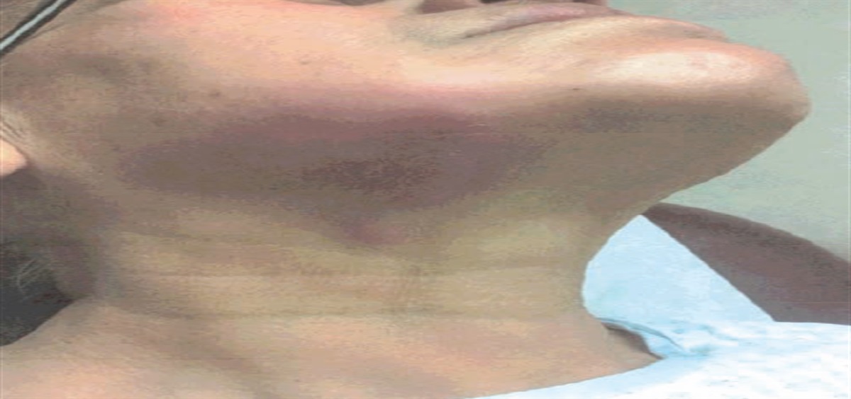
CLINICAL PRESENTATIONThe clinical presentation of MCC typically is described as a firm, painless violaceous nodule that can ulcerate or become hyperkeratotic as it increases in size and that is associated with telangiectasia or a shiny surface (Figure 1).29,30 Early MCC lesions can look like a cyst or acneiform lesion, a vascular lesion, an insect bite, or a subcutaneous nodule (Table 1).31 Other less common features include a diffuse bruise-like vascular pattern, milky red patches, and disconnected branching vessels.32 Patients with advanced stages of MCC may develop lymphadenopathy.30 Because of the nonspecific clinical findings, MCC cannot be diagnosed without a biopsy. However, literature has shown that 89% of MCC is characterized by at least three of the following symptoms in the mnemonic AEIOU: asymptomatic/lack of tenderness, expanding rapidly, immunosuppression, older than age 50 years, and UV-exposed site on a person with fair skin.31
 FIGURE 1.:
 An erythematous dome-shaped nodule (2.2x1.5cm) on the upper aspect of the right shoulder of a 72-year-old woman. Photograph courtesy of Jo Bohannon-Grant, MD
TABLE 1. - Clinical differential diagnosis of MCC58-65 Clinical presentation Histologic presentation Epidermoid cyst Skin-colored, mobile nodule with or without a central punctum The lining along the epithelial cells lacks rete pegs. The region contained by the lining has keratin along with a granular layer of keratin granules. BCC Variable presentations. Nonhealing erosions, erythematous papule with telangiectasia and raised pearly borders. Aggregation of basophilic cells surrounded by stromal cells. Keratinocytes contain hyperchromatic nuclei with smooth or coarse chromatin. Melanoma Variation presentation. Flat, dark-pigmented network/patch with color variation, erythematous nodule, or cerebriform appearance. May be associated with inconsistent border or ulceration. The cells lining the epidermis layer shows ulceration, frequent mitosis, and lack of maturation. The cells can grow solitarily or in aggregates. Cutaneous lymphoma Variable presentation. Erythematous patches, disseminated nodules, or ulcerated plaques. May be associated with itchiness and lymphadenopathy. Variable histopathology. Most contain intraepidermal lymphocyte aggregation and angiocentricity. Cells are mononuclear and exhibit a sheet growth pattern. Fibroxanthoma Asymptomatic, ulcerating nodule No specific defining histologic features. Variants may include nonpleomorphic spindle cells, keloidal cells, or granular cells.On histopathology, MCC appears as densely packed blue cells containing granular chromatin, also known as the salt-and-pepper chromatin (Figures 2 and 3).33 MCC cells typically are in nests and have minimal cytoplasm. On lower magnification, numerous mitotic figures and central necrotic regions also can be observed.33,34 To confirm the diagnosis of MCC, immunohistochemical stains are used. MCC usually is positive for cytokeratin 20 (CK20), transcription factor special AT-rich sequence-binding protein 2, neurofilament, and MCV.35 Immunohistochemical stains also help differentiate it from other mimics.
 FIGURE 2.:
 MCC variant showing a collection of dermal and subcutaneous cells with basophilic nuclei with high mitotic activity at 2.5 times magnification
 FIGURE 3.:
 MCC variant showing cells with basophilic nuclei with high mitotic activity at 40 times magnification
DIFFERENTIAL DIAGNOSISThe clinical differential diagnosis of MCC includes epidermal inclusion cyst, BCC, amelanotic melanoma, lymphoma, or atypical fibroxanthoma (Table 1).36
STAGINGAccording to the AJCC Cancer Staging Manual, 8th ed., from the American Joint Committee on Cancer, MCC staging depends on tumor size (T), lymph node invasion (N), and the extent of metastasis to other organs (M), characteristics used to classify tumors from stage 1 to stage 4.37 The classification of clinical versus pathologic N stage corresponds with stage-specific survival rate, with patients with positive lymph node invasion having a lower survival rate.38
Primary tumor: T stageTumors that cannot be directly measured are classified as Tx. Tumors that cannot be found on site are classified as T0, and those in situ are classified as Tis. For tumors that have spread beyond the submucosa, the staging is dictated by their diameter. Tumors 2 cm or smaller are classified as T1, or stage 1. Tumors larger than 2 cm but smaller than 5 cm are classified as T2, or stage 2. Tumors 5 cm or larger are classified as stage 3 or stage 4, with the difference being that stage 4 tumors have evidence of involving other organs.37
Regional lymph nodes: N stageNegative lymph node involvement is classified as the clinical stage and positive lymph node involvement as the pathologic stage. N1A is defined as tumor size between 0.2 mm and 2 mm in the lymph nodes; any tumor size beyond 2 mm in the lymph nodes is defined as N1B.39 However, tumors with evidence of in-transit metastasis in the lymph nodes are classified as N2.39 Only stage 3 and stage 4 tumors have evidence of lymph node invasion.37 Stage 3A is defined as a positive lymph node identified by pathology; stage 3B is positive identification via radiology or other clinical evidence.40
Distant metastasis: M stageOnly stage 4 tumors have evidence of organ metastasis.37 M1A is defined as metastasis to skin, subcutaneous regions, and distant lymph nodes.39 M1B is evidence of metastasis to the lung, and M1C is metastasis to other visceral organs beyond the lungs.39
DIAGNOSISIf a patient has lesions suspicious for MCC, perform a complete body and skin examination followed by lesion biopsy.41 Fine-needle aspiration cytology is a common, noninvasive method to obtain samples from the lesion.42 The biopsied sample should then undergo hematoxylin and eosin staining, followed by immunohistochemistry (IHC) staining for confirmation.41 The antibodies used in IHC staining should include CK20 and thyroid transcription factor-1 at minimum, but other markers can be included.41 The characteristics of the lesion should be evaluated by an experienced pathologist, who can provide adequate information on tumor thickness, mitotic rate, growth pattern, infiltrating lymphocytes, and other malignancies (Figure 4).41,43 Because of MCC's metastatic potential and the possibility of MCC coexisting with other tumors, obtain biopsies of different lesions.41
 FIGURE 4.:
 MCC growth pattern: nodular growth pattern (a, low magnification; b, high magnification) and infiltrative growth pattern (c, low magnification; d, high magnification)Reproduced with permission from Andea AA, Coit DG, Amin B, Busam KJ. Merkel cell carcinoma: histologic features and prognosis. Cancer. 2008;113(9):2549-2558.
Obtain sentinel lymph node biopsy (SLNB) in all patients with MCC to detect possible nodal spread (National Comprehensive Cancer Network [NCCN] evidence category 2A).44 If evidence of lymph node involvement is identified, obtain further imaging. Fluorodeoxyglucose (FDG)-positron emission tomography (PET)/CT scanning is the most common imaging method for patients with MCC, but if it is unavailable, CT scan or MRI with contrast may be used.41 Obtain whole-body imaging if the patient has evidence of distant metastasis.41 In patients with evidence of brain, spine, or bone metastasis, obtain a focused MRI with contrast.41,45
Patients with positive SLNBs should undergo nodal dissection and/or adjuvant radiotherapy, which requires referral to a radiation oncologist.46 In patients with advanced stages of MCC, a medical oncologist also is needed to guide chemotherapy (NCCN evidence category 2A).46
TREATMENTFor patients who cannot undergo or decline surgical excision, radiotherapy alone is recommended.47 After excision, observation of the region may be preferred over radiotherapy, especially during early stages of MCC.47
Patients diagnosed with stage 2 MCC also undergo surgical excision of the affected region with 1- to 2-cm margins, followed by radiotherapy.47 Mohs micrographic surgery can be used instead of surgical excision when MCC occurs on the head and neck, and can be performed after SLNB to prevent damage to the draining lymph nodes.48
Patients with positive results from SLNB are classified as stage 3, which suggests that they will need a PET/CT scan to examine the extent of the metastasis.47 Imaging also should be performed on all high-risk patients and those in whom aggressive clinical features are noted.49 If no evidence of further metastasis to other organs is found, lymph node dissection followed by radiotherapy is recommended.47
Patients with evidence of further metastasis to other organs are classified as stage 4. They can either undergo radiotherapy, immunotherapy, or chemotherapy. The benefits of chemotherapy on survival rate only last for about 3 months; more research is pointing to immunotherapy because of its durable effect.48,50 Avelumab is one of the FDA-approved immunotherapy drugs for MCC and only is used to treat advanced stages.50 Patients who are not candidates for immunotherapy because of pregnancy or autoimmune disease are treated with chemotherapy drugs, with platinum and etoposide being among the most common agents.47,51
According to the NCCN, patients should have follow-up visits every 3 to 6 months for 3 years, and every 6 to 12 months afterwards. This follow-up should involve a physical examination with complete skin and lymph node examination.47 Consider more frequent follow-up for high-risk patients, such as older adults and those with immunosuppression.47 MCV-oncoprotein antibodies are a good marker for screening recurrence rate, with a 66% positive predictive value of elevated antibody titers associated with higher recurrence rate.52 If recurrence occurs, different measures are taken depending on the extent of metastasis. If metastasis only occurs in a localized region, the affected region may undergo resection when possible. If recurrence results in lymph node invasion, the patient may undergo lymph node dissection. Consider chemotherapy for patients with primary or recurrent MCC that involves metastasis beyond lymph node invasion.53
CONCLUSIONAlthough MCC is rare, early diagnosis and treatment are key to successful management. Physician associates/assistants (PAs) should be aware of typical and uncommon presentations of MCC and maintain a high level of scrutiny when examining each skin lesion, no matter how similar they present to more common, benign skin findings. When presentation is unclear, biopsy with histopathologic staining should be performed to differentiate between MCC and other cutaneous skin lesions.
PAs also play a crucial role in educating patients and their families on the risks and benefits of each therapeutic option, as well as the consequences of not pursuing treatment. In cases where radiotherapy or chemotherapy is included as part of the regimen, studies have shown that preparatory information that addresses adverse reactions to these treatments, such as vomiting, fatigue, hair loss, and sleep pattern changes, can lead to better psychosocial outcomes and greater patient satisfaction.54
Counsel all patients on skin cancer preventive measures, such as reduction of sun exposure, application of broad-spectrum sunscreen with a sun protection factor of 30 or higher, and avoidance of tanning beds.55 Educate patients about vaccine development to target MCV capsid proteins VP1 and VP2, although further testing is needed before these vaccines are made available to the public.56,57
REFERENCES 1. Woo S-H, Lumpkin EA, Patapoutian A. Merkel cells and neurons keep in touch. Trends Cell Biol. 2015;25(2):74–81. 2. Hoffman BU, Baba Y, Griffith TN, et al. Merkel cells activate sensory neural pathways through adrenergic synapses. Neuron. 2018;100(6):1401–1413. 3. Wright MC, Logan GJ, Bolock AM, et al. Merkel cells are long-lived cells whose production is stimulated by skin injury. Dev Biol. 2017;422(1):4–13. 4. American Cancer Society. Key statistics for Merkel cell carcinoma. www.cancer.org/cancer/merkel-cell-skin-cancer/about/key-statistics.html. Accessed August 7, 2023. 5. American Cancer Society. Basal and squamous cell skin cancer statistics. www.cancer.org/cancer/basal-and-squamous-cell-skin-cancer/about/key-statistics.html. Accessed August 7, 2023. 8. American Cancer Society. Survival rates for Merkel cell carcinoma. www.cancer.org/cancer/merkel-cell-skin-cancer/detection-diagnosis-staging/survival-rates.html. Accessed August 7, 2023. 9. Piva de Freitas P, Senna CG, Tabai M, et al. Metastatic basal cell carcinoma: a rare manifestation of a common disease. Case Rep Med. 2017;2017:8929745. 10. Shreve C, Shropshire C, Cotter DG. Metastatic squamous cell carcinoma: a cautionary tale. Cureus. 2020;12(10):e10879. 11. Sandru A, Voinea S, Panaitescu E, Blidaru A. Survival rates of patients with metastatic malignant melanoma. J Med Life. 2014;7(4):572–576. 12. Lewis CW, Qazi J, Hippe DS, et al. Patterns of distant metastases in 215 Merkel cell carcinoma patients: implications for prognosis and surveillance. Cancer Med. 2020;9(4):1374–1382. 13. Becker JC, Stang A, DeCaprio JA, et al. Merkel cell carcinoma. Nat Rev Dis Primers. 2017;3:17077. 14. Harms PW, Harms KL, Moore PS, et al. The biology and treatment of Merkel cell carcinoma: current understanding and research priorities. Nat Rev Clin Oncol. 2018;15(12):763–776. 15. Sihto H, Kukko H, Koljonen V, et al. Merkel cell polyomavirus infection, large T antigen, retinoblastoma protein and outcome in Merkel cell carcinoma. Clin Cancer Res. 2011;17(14):4806–4813. 16. Tolstov YL, Pastrana DV, Feng H, et al. Human Merkel cell polyomavirus infection II. MCV is a common human infection that can be detected by conformational capsid epitope immunoassays. Int J Cancer. 2009;125(6):1250–1256. 17. Verhaegen ME, Mangelberger D, Harms PW, et al. Merkel cell polyomavirus small T antigen initiates Merkel cell carcinoma-like tumor development in mice. Cancer Res. 2017;77(12):3151–3157. 18. Baez CF, Brandão Varella R, Villani S, Delbue S. Human polyomaviruses: the battle of large and small tumor antigens. Virology (Auckl). 2017;8:1178122X17744785. 19. Houben R, Adam C, Baeurle A, et al. An intact retinoblastoma protein-binding site in Merkel cell polyomavirus large T antigen is required for promoting growth of Merkel cell carcinoma cells. Int J Cancer. 2012;130(4):847–856. 20. Cornejo C, Miller CJ. Merkel cell carcinoma: updates on staging and management. Dermatol Clin. 2019;37(3):269–277. 21. Silling S, Kreuter A, Gambichler T, et al. Epidemiology of Merkel cell polyomavirus infection and Merkel cell carcinoma. Cancers (Basel). 2022;14(24):6176. 22. Ma JE, Brewer JD. Merkel cell carcinoma in immunosuppressed patients. Cancers (Basel). 2014;6(3):1328–1350. 23. Izikson L, Nornhold E, Iyer JG, et al. Merkel cell carcinoma associated with HIV: review of 14 patients. AIDS. 2011;25(1):119–121. 24. Clarke CA, Robbins HA, Tatalovich Z, et al. Risk of Merkel cell carcinoma after solid organ transplantation. J Natl Cancer Inst. 2015;107(2):dju382. 25. Cancer.Net. Skin cancer (non-melanoma)—risk factors and prevention. www.cancer.net/cancer-types/skin-cancer-non-melanoma/risk-factors-and-prevention. Accessed August 7, 2023. 26. Guadagni S, Farina AR, Cappabianca LA, et al. Multidisciplinary treatment, including locoregional chemotherapy, for Merkel-polyomavirus-positive Merkel cell carcinomas: perspectives for patients exhibiting oncogenic alternative Δ exon 6-7 TrkAIII splicing of neurotrophin receptor tropomyosin-related kinase A. Int J Mol Sci. 2020;21(21):8222. 27. Ursu RG, Damian C, Porumb-Andrese E, et al. Merkel cell polyoma virus and cutaneous human papillomavirus types in skin cancers: optimal detection assays, pathogenic mechanisms, and therapeutic vaccination. Pathogens. 2022;11(4):479. 28. Hafner C, Houben R, Baeurle A, et al. Activation of the PI3K/AKT pathway in Merkel cell carcinoma. PLoS ONE. 2012;7(2):e31255. 29. Agni M. Dermatologic manifestations of Merkel cell carcinoma clinical presentation: history, physical examination, complications. https://emedicine.medscape.com/article/1100917-clinical. Accessed August 7, 2023. 30. National Organization for Rare Disorders. Merkel cell carcinoma. https://rarediseases.org/rare-diseases/merkel-cell-carcinoma. Accessed August 7, 2023. 31. Heath M, Jaimes N, Lemos B, et al. Clinical characteristics of Merkel cell carcinoma at diagnosis in 195 patients: the AEIOU features. J Am Acad Dermatol. 2008;58(3):375–381. 32. Laureano A, Cunha D, Pernandes C, Cardoso J. Dermoscopy in Merkel cell carcinoma: a case report. Dermatol Online J. 2014;20(2). 33. Pulitzer M. Merkel cell carcinoma. Surg Pathol Clin. 2017;10(2):399–408. 34. Bailey TL, Fung MA, Gandour-Edwards R, et al. Clinical emergence of neurometastatic Merkel cell carcinoma: a surgical case series and literature review. J Neurooncol. 2011;102(1):147–155. 35. Kervarrec T, Tallet A, Miquelestorena-Standley E, et al. Diagnostic accuracy of a panel of immunohistochemical and molecular markers to distinguish Merkel cell carcinoma from other neuroendocrine carcinomas. Mod Pathol. 2019;32(4):499–510. 36. Jaeger T, Ring J, Andres C. Histological, immunohistological, and clinical features of Merkel cell carcinoma in correlation to Merkel cell polyomavirus status. J Skin Cancer. 2012;2012:983421. 37. Wang TS, Byrne PJ, Jacobs LK, Taube JM. Merkel cell carcinoma: update and review. Semin Cutan Med Surg. 2011;30(1):48–56. 38. Allen PJ, Bowne WB, Jaques DP, et al. Merkel cell carcinoma: prognosis and treatment of patients from a single institution. J Clin Oncol. 2005;23(10):2300–2309. 39. Petrov A, Kraleva S, Kubelka-Sabit K, Petrova D. Treatment of a patient with Merkel cell skin carcinoma using radiation therapy—a case report. Open Access Maced J Med Sci. 2018;6(4):669–672. 40. Hughes MP, Hardee ME, Cornelius LA, et al. Merkel cell carcinoma: epidemiology, target, and therapy. Curr Dermatol Rep. 2014;3(1):46–53. 41. Bichakjian CK, Olencki T, Aasi SZ, et al. Merkel cell carcinoma, Version 1.2018, NCCN Clinical Practice Guidelines in Oncology. J Natl Compr Canc Netw. 2018;16(6):742–774. 42. Tai P. A practical update of surgical management of Merkel cell carcinoma of the skin. ISRN Surg. 2013;2013:850797. 43. Andea AA, Coit DG, Amin B, Busam KJ. Merkel cell carcinoma: histologic features and prognosis. Cancer. 2008;113(9):2549–2558. 44. Gupta SG, Wang LC, Peñas PF, et al. Sentinel lymph node biopsy for evaluation and treatment of patients with Merkel cell carcinoma: the Dana-Farber experience and meta-analysis of the literature. Arch Dermatol. 2006;142(6):685–690. 45. Tirumani SH, Shinagare AB, Sakellis C, et al. Merkel cell carcinoma: a primer for the radiologist. AJR. 2013;2000(6):1186–1196. 46. Prieto I, Pérez de la Fuente T, Medina S, et al. Merkel cell carcinoma: an algorithm for multidisciplinary management and decision-making. Crit Rev Oncol Hematol. 2016;98:170–179. 47. Schmults C, Blitzblau R, Aasi S, et al. NCCN clinical practice guidelines in oncology for Merkel cell carcinoma. National Comprehensive Cancer Network. 2021. https://merkelcell.org/wp-content/uploads/2022/04/NCCN-Guidelines-for-Merkel-Cell-Carcinoma-v1.2023.pdf. Accessed August 7, 2023. 48. Burcoş T, Popa E, Ivăşcanu A, et al. Carcinomul cu celule Merkel [Merkel cell carcinoma]. Chirurgia (Bucur). 2001;96(5):509–516. 49. Cassler NM, Merrill D, Bichakjian CK, Brownell I. Merkel cell carcinoma therapeutic update. Curr Treat Options Oncol. 2016;17(7):36. 50. Baker M, Cordes L, Brownell I. Avelumab: a new standard for treating metastatic Merkel cell carcinoma. Expert Rev Anticancer Ther. 2018;18(4):319–326. 51. Banks PD, Sandhu S, Gyorki DE, et al. Recent insights and advances in the management of Merkel cell carcinoma. J Oncol Pract. 2016;12(7):637–646. 52. Paulson KG, Lewis CW, Redman MW, et al. Viral oncoprotein antibodies as a marker for recurrence of Merkel cell carcinoma: a prospective validation study. Cancer. 2017;123(8):1464–1474. 53. Beenken SW, Urist MM. Treatment options for Merkel cell carcinoma. J Natl Compr Canc Netw. 2004;2(1):89–92. 54. Waller A, Forshaw K, Bryant J, Mair S. Interventions for preparing patients for chemotherapy and radiotherapy: a systematic review. Support Care Cancer. 2014;22(8):2297–2308. 55. Pérez LL, Bashline B. Skin cancer: prevention. FP Essent. 2019;481:28–31. 56. Xu D, Jiang S, He Y, et al. Development of a therapeutic vaccine targeting Merkel cell polyomavirus capsid protein VP1 against Merkel cell carcinoma. NPJ Vaccines. 2021;6(1):119. 57. Almansour NM. Immunoinformatics- and bioinformatics-assisted computational designing of a novel multiepitopes vaccine against cancer-causing Merkel cell polyomavirus. Front Microbiol. 2022;13:929669. 58. Zito PM, Scharf R. Epidermoid cyst. Treasure Island, FL: StatPearls Publishing; 2020. www.ncbi.nlm.nih.gov/books/NBK499974. Accessed August 7, 2023. 59. McCourt C, Dolan O, Gormley G. Malignant melanoma: a pictorial review. Ulster Med J. 2014;83(2):103–110. 60. Marzuka AG, Book SE. Basal cell carcinoma: pathogenesis, epidemiology, clinical features, diagnosis, histopathology, and management. Yale J Biol Med. 2015;88(2):167–179. 61. Tetzlaff MT, Nagarajan P. Update on Merkel cell carcinoma. Head Neck Pathol. 2018;12(1):31–43. 62. Filosa A, Filosa G. Melanoma diagnosis: the importance of histopathological report. Dermatopathology (Basel). 2018;5(1):41–43. 63. Kempf W, Zimmermann A-K, Mitteldorf C. Cutaneous lymphomas—an update 2019. Hematol Oncol. 2019;37(suppl 1):43–47. 64. Slater D. Histopathological aspects of cutaneous lymphoma. J R Soc Med. 2001;94(7):337–340. 65. Sarac E, Yuksel M, Turkmen IC, Ozdemir M. Case for diagnosis. Atypical fibroxanthoma. An Bras Dermatol. 2019;94(2):239–241.
Comments (0)