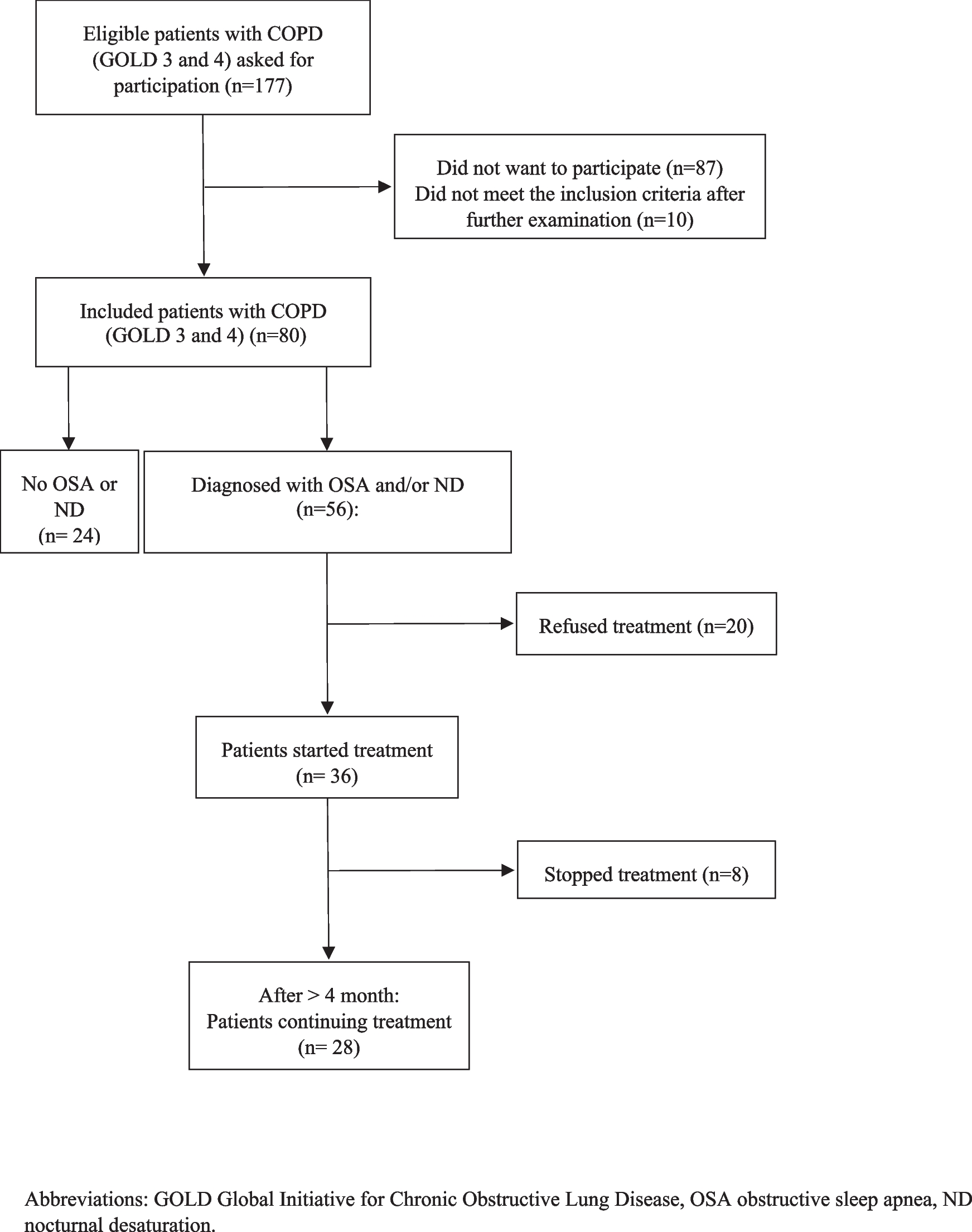
Precise identification of the obstruction site in patients with OSA is an important initial step towards achieving successful surgical outcomes [25]. Awake endoscopy with MM and drug-induced sleep endoscopy (DISE) both provide a topographical evaluation of the upper airway. Both maneuvers help sleep surgeons in evaluating the level and the pattern of upper airway collapse. Currently, awake endoscopy with MM and DISE have become the most reliable tools in evaluating upper airway collapsibility of patients with OSA. However, both methods have their inherent limitations [26,27,28]. MM is an outpatient procedure that can be performed without drug induction. MM is less burdensome on the patient and hospital. However, controversy exists regarding the accuracy of evaluating upper airway collapsibility grade and pattern during MM due to its performance on awake patients. Due to its performance during sleep, DISE is widely regarded by sleep surgeons as the most dependable tool for assessing upper airway collapsibility. However, DISE has its limitations. DISE requires trained personnel, specialized facilities, and special rooms for the procedure. Additionally, the drug-induced sleep achieved during DISE only captures a limited portion of the complete sleep cycle, potentially failing to reflect all changes in upper airway morphology throughout the entire cycle. Furthermore, sleep surgeons encounter challenges in conducting follow-up assessments of postoperative patients using DISE due to its high cost and the requirement for general anesthesia. Consequently, DISE is not extensively utilized, particularly in low-income countries with restricted healthcare budgets.
Previous research has compared the outcomes of DISE and MM, revealing that both tests can significantly affect surgical planning and decision-making by providing precise assessments of upper airway collapse patterns and levels [3,4,5, 24,25,26,27,28,29]. Recent findings indicate that the obstruction pattern observed in DISE and MM is comparable, with the degree of collapse being the distinguishing factor [20, 30, 31]. Askar et al. demonstrated that positional awake endoscopy is a cost-effective and convenient outpatient procedure that yields comparable results to DISE in terms of upper airway collapse patterns and grades at all levels [20]. Unlike DISE, supine MM does not necessitate specialized anesthesia precautions, equipment, or facilities. The follow-up of patients utilizing supine MM is significantly simplified, and the issue of patient consent can be more readily resolved. Overall, the supine MM can provide valuable surgical insights into the level, pattern, and degree of upper airway collapse in patients with OSA. Therefore, we consider the supine MM to be a reliable tool for evaluating the three-dimensional anatomical topography of the upper airway and for making informed surgical plans and decisions.
The retrolingual obstruction is often observed in patients with severe OSA [4,5,6]. In order to minimize trauma, the transoral approach is typically preferred when treating such patients. Alternative tools include laser treatment, radiofrequency, and transoral robotic surgery. In recent years, otolaryngologists have increasingly utilized the coblation technique, which represents a cutting-edge and innovative approach to surgical procedures. Energized electrodes immersed in saline solution generate a plasma layer consisting of highly ionized particles, which effectively disrupt intercellular bonds within tissue and can be removed at low temperatures. Compared to other approaches, coblation is associated with reduced morbidity and fewer complications.
In 2006, Maturo and Mair first performed tongue base resection via coblation [32]. Li et al. pioneered coblation endoscopic lingual lightening for patients with OSA due to retrolingual obstructions [11]. The central portion of a hypertrophic tongue base is transorally ablated using coblation under endoscopic guidance. Bahgat et al. have introduced a novel transoral tongue base surgery technique, referred to as the “Robo-Cob” technique [9]. The exposure and operative technique are analogous to those of transoral robotic surgery. However, coblation is utilized for tongue base tissue resection instead of ablation. Additional coblation techniques have also been delineated [10, 14, 33, 34], all of which are viable and moderately effective in addressing retrolingual obstructions. However, the surgical response rates were suboptimal and postoperative morbidity and complications, including pain and bleeding, were relatively prevalent.
Previous treatments for retrolingual obstruction have primarily focused on the tongue base, neglecting the importance of the tongue body [35]. However, it is important to note that both the tongue base and body can contribute to not only retrolingual obstruction but also retropalatal obstruction [6, 36, 37]. Both regions are capable of causing retrolingual obstruction by moving against the posterior pharyngeal wall, and can also induce retropalatal obstruction by pushing the soft palate back against it. In clinical practice, the majority of patients with severe OSA exhibit hypertrophy of the tongue body, making treatment of this area a crucial aspect in managing their condition. The hypoglossal neurovascular bundle is situated at a depth greater than 1.5 cm and blood supply to the middle region of the tongue body is relatively poor [38]. Therefore, the surgical scope of tongue base and tongue body in our study was deemed safe. However, there is no standardized approach for managing the tongue body during treatment of retrolingual obstruction, and it remains unclear how much tissue can be safely resected to achieve optimal postoperative outcomes while minimizing functional impairment. In the current study, the ablation of the tongue body commenced 3–4 cm anterior to the circumvallate papillae and subsequently extended posteriorly along the midline towards the base of the tongue. The ablated region measured 2 cm in width and 1.5 cm in depth. Endoscopy findings revealed a significant increase in the anteroposterior diameter, transverse diameters, and cross-sectional area of the retrolingual region postoperatively.
The surgical response rates of these tongue base procedures exhibit significant variability, ranging from 56.3 to 78.7% [1, 8, 10, 11, 13, 39]. The current study demonstrated significant improvement in surgical response rates compared to previous research. Furthermore, at the 6-month follow-up, no lingual dysfunction was reported, indicating that our lingual surgery appears to be both safe and effective.
Previous research has suggested that the inclusion of lingual surgery in pharyngoplasty procedures may increase the likelihood of morbidity and complications, such as bleeding, pain, infection, edema, lingual paralysis, and taste disturbance [10, 11, 13, 18]. However, our own experience did not support this conclusion. The lingual ablated area was left unsutured in previous studies, whereas we opted to close the same using 2–0 absorbable threads. Lee et al. conducted a meta-analysis of nine studies on the use of plasma ablation for tongue base reduction in patients with OSA, revealing a 7.5% incidence rate of postoperative bleeding [12]. Sutures were not conduced in previous tongue base studies. In the current study, only one case of postoperative bleeding was encountered, indicating a low likelihood of such an occurrence. The reasons are twofold. First, suturing can occlude and ligate damaged blood vessels. Second, the ablated region lacking sutures induces the formation of protective pseudo-membranes that envelop the wound site and gradually dissolve during the process of healing. Premature displacement of these membranes and hemorrhage may occur due to early consumption of solid or hot foods, wound infection, and frequent coughing caused by abnormal throat sensations. Therefore, sutures can close the surgical wound, accelerate healing, and reduce postoperative bleeding.
There are several limitations to the study that need to be addressed. First, DISE was not utilized for the evaluation of the upper airway. Second, the efficacy of modified tongue surgery in multilevel surgery may be affected by the concurrent effect of H-UPPP. Therefore further investigation is required with further study. Third, the limited sample size utilized in this study restricted the extent of analysis regarding factors associated with outcomes. Additionally, a more extended follow-up period would facilitate the identification of ideal candidates for modified coblation endoscopic lingual lightening treatment.

Comments (0)