
Remember me


Pyoderma gangrenosum (PG) is a rare inflammatory skin disorder in the category of neutrophilic dermatoses that exists in various subtypes. Postoperative PG (PPG) is an uncommon subtype that occurs approximately 2 weeks after surgery.1 Postoperative PG is most commonly observed after breast surgery, thoracic and cardiac surgery, and abdominal surgery.
CASE REPORTA 55-year-old Chinese man presented to the hospital with abdominal pain and distension for 1 month. The patient had an unremarkable medical history. He was diagnosed with small bowel obstruction and underwent partial laparoscopic small bowel resection, after which a pelvic drain was placed. No stoma was created. Pathologic examination of the small intestine showed that the mucosal layer of the intestinal wall had inflammatory granulation tissue formation, and the submucosal layer had interstitial edema with inflammation (Figure 1).
 Figure 1.:
 PATHOLOGY OF SMALL INTESTINE BIOPSYThe mucosal layer of the intestinal wall had inflammatory granulation tissue formation; the submucosal layer had interstitial edema with inflammation (hematoxylin-eosin stain, original magnification ×100).
On postoperative day 6, the patient developed a fever of approximately 38.5 °C, and his blood count indicated elevated levels of leukocytes (16 × 109/L; reference range, 3.5-9.5 × 10⁹/L) and neutrophils (14 × 109/L; reference range, 1.8-6.3 × 10⁹/L). Providers also noted redness and bleeding from the incision (Figure 2) and yellow-green turbid drainage fluid. An incisional secretion culture revealed Pseudomonas aeruginosa (+), and the blood culture was negative. The clinician suspected postoperative skin infection, and the patient was treated with antibiotics (imipenem, 1000 mg every 8 hours). The patient also received continuous abdominal irrigation with double trocars, daily dressing changes, and oil gauze after wound cleaning without improvement.
 Figure 2.:
 WOUND CONDITION ON POSTOPERATIVE DAY 6The median abdominal incision was red and swollen with oozing blood. The edge was irregular, and the skin around the two tube orifices was red.
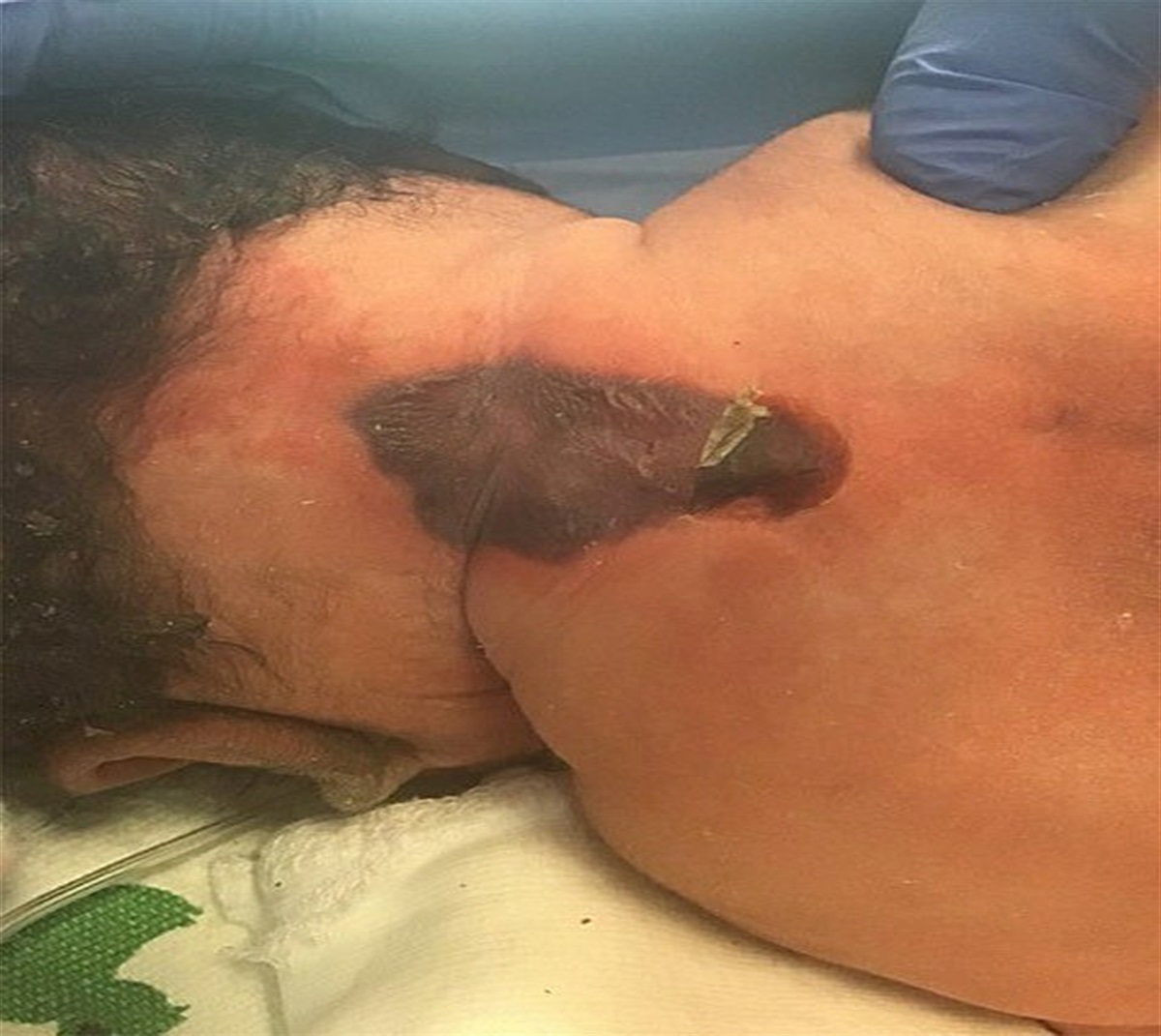
On postoperative day 7, the provider observed a 2 × 2-cm erythema in the median abdominal incision with blistered edges that bled easily after rupture. The patient reported that the pain was unbearable. A dermatologist was consulted. On physical examination, which was led by the dermatologist with joint participation by general surgeons and nurses, three ulcers were found in the middle of the abdomen. The largest ulcer was 10 × 10 cm and irregularly shaped, with undermined wound edges; surrounding violaceous, blue-gray erythema (Figure 3); and obvious tenderness. Therefore, the possibility of PPG was considered and improved histopathology, and multiple secretion cultures were performed. The patient was also screened for systemic diseases, including autoantibody, humoral immunity, tumor marker, rheumatoid factor, and thyroid function assessments. After 3 days, pathology revealed many neutrophil infiltrates and abscess formation in the dermis, with focal epidermal rupture and ulcer formation (Figure 4).
 Figure 3.:
 WOUND CONDITION ON POSTOPERATIVE DAY 10An irregular ulcer measuring 10 × 10 cm developed in the middle of the abdomen, with undermined wound edges and surrounding violaceous, blue-gray erythema. A crater-like depression can be seen in the center. Two small, irregular ulcers, which measured 2 × 2 cm and 3 × 3 cm, developed around the tubes, with surrounding violaceous, blue-gray erythema. The surface was covered with pseudomembranous substances and purulent exudate.
 Figure 4.:
 WOUND BIOPSYFocal epidermal rupture with ulcer formation and a large number of neutrophil infiltrates and abscess formation in the dermis (hematoxylin-eosin stain; original magnification ×100).
The patient was treated with systemic corticosteroids (methylprednisolone sodium succinate 40 mg/d) combined with intravenous immunoglobulin (20 g/d, 3 days); antibiotics were adjusted according to the patient’s drug allergies to meropenem, ceftazidime, cefoperazone/sulbactam, and amikacin. The patient was also administered a wet compress of Kangfuxin solution for 30 minutes daily, and the area was covered with a nonadherent dressing. After 1 week of treatment, the patient’s condition improved: his fever abated, the ulcer area did not expand, exudation decreased on the day after hormone treatment, and the levels of leukocytes and neutrophils returned to normal. Two weeks later, the patient gradually switched to a semiliquid diet, and his appetite improved.
The patient provided written informed consent for publication of the case details and images.
DISCUSSIONPostoperative PG is an independent subtype of PG that manifests following surgical incision or trauma, generally occurring within 2 weeks of surgery and averaging 7 days.1 The pathogenesis of PPG is unknown and may be related to the release of inflammatory mediators from cellular activation caused by surgical incision irritation or traction. The earliest clinical sign of PPG is erythema around the incision sutures. Subsequently, the wound may form small areas of discharge with punctate ulcerations that coalesce into larger areas of wound dehiscence.2 In contrast to other forms of PG, PPG has a predictable onset time, occurs most often on the trunk, and has a low correlation with systemic diseases.3 Most patients experience antibiotic treatment and debridement at the onset. Therefore, PPG remains a diagnosis of exclusion that requires the elimination of other causes of skin ulcers, such as primary infection, malignancy, and vasculitis.4 In real-world practice, coinfection with PG is common. In a multicenter study, Binus et al5 found colonization by one or more bacterial colonies in the tissue samples of a considerable proportion of patients with PG.5 Further, an infection can be an environmental trigger or an additional factor in PG development.
In this case, the patient presented at disease onset with edematous erythema and rapid progression to a necrolytic ulcer within 3 days, according to the typical manifestations of PG. Although the secretion culture was positive during this period, antibiotics and debridement treatment were ineffective, and the diagnosis of PPG was considered in combination with histopathologic findings. In this case, the secretion cultures suggesting positive P aeruginosa were considered secondary bacterial infections. Therefore, in such situations, doctors should select sensitive antibiotics at a full dose; however, if ineffective, the cause of the ulcer should be actively investigated.
The systemic use of glucocorticoid is still the first-line treatment for PG, and combined immunosuppressant usage can reduce the glucocorticoid dose.6 In addition, because antitumor necrosis factor drugs such as infliximab and adalimumab are safe and effective at treating neutrophil-related diseases, they may also have good efficacy in PG treatment, particularly in patients with inflammatory bowel disease (IBD).7 Dowd et al8 reported that the prophylactic use of infliximab in patients with PPG requiring surgical intervention reduces the risk of recurrence.8 Because PG can require surgical treatment, including split-thickness skin grafts and vascularized free tissue transfer,9 immunosuppressive agent use during this period helps avoid disease recurrence and deterioration.
In the present case, the providers considered the nature of the relationship between PPG and the gastrointestinal tract because the patient had an acute onset following gastrointestinal symptoms that required surgical treatment. As a systemic disease, PG can affect the digestive system, and its relationship with IBD has been the most studied. Known relationships between PG and the intestine include: (1) PG with multiple organ involvement involves the gastrointestinal tract, (2) PG is associated with IBD,10 and (3) PG is a cutaneous manifestation of IBD.11 The relationship between PG and intestinal lesions deserves deeper consideration. In general, PG with IBD or PG as a cutaneous manifestation of IBD follows a certain time sequence: skin manifestations usually appear in the active/poorly controlled phase of the original disease, which was inconsistent with the patient described in this report. Further, no evidence of systemic involvement of the conjunctiva, cornea, oral mucosa, or lungs was found, decreasing the likelihood of multiple organ involvement in this patient.
CONCLUSIONSPostoperative PG and intestinal symptoms have various complex relationships and should be considered comprehensively based on the patient’s medical history and clinical presentation. In clinical practice, patient outcomes are improved with early identification, avoidance of unnecessary debridement, and timely selection of reasonable immunosuppressive therapies.
REFERENCES 1. Tolkachjov SN, Fahy AS, Wetter DA, et al. Postoperative pyoderma gangrenosum (PG): the Mayo Clinic experience of 20 years from 1994 through 2014. J Am Acad Dermatol 2015;73:615–22. 2. Ogon M, Sepp NT, Wimmer C, Behensky H. A surgical wound infection? Lancet 2000;356:1652. 3. Tolkachjov SN, Fahy AS, Cerci FB, Wetter DA, Cha SS, Camilleri MJ. Postoperative pyoderma gangrenosum: a clinical review of published cases. Mayo Clin Proc 2016;91:1267–79. 4. Su WPD, Davis MDP, Weenig RH, Powell FC, Perry HO. Pyoderma gangrenosum: clinicopathologic correlation and proposed diagnostic criteria. Int J Dermatol 2004;43:790–800. 5. Binus AM, Qureshi AA, Li VW, Winterfield LS. Pyoderma gangrenosum: a retrospective review of patient characteristics, comorbidities and therapy in 103 patients. Br J Dermatol 2011;165:1244–50. 6. Zuo KJ, Fung E, Tredget EE, Lin AN. A systematic review of post-surgical pyoderma gangrenosum: identification of risk factors and proposed management strategy. J Plast Reconstr Aesthet Surg 2015;68:295–303. 7. Hubbard VG, Friedmann AC, Goldsmith P. Systemic pyoderma gangrenosum responding to infliximab and adalimumab. Br J Dermatol 2005;152:1059–61. 8. Dowd M, Samie F, Gallitano S. Infliximab prophylaxis in patients with postoperative pyoderma gangrenosum requiring surgical intervention. Dermatol Surg 2021;47:272–74. 9. Bingoel AS, Krezdorn N, Kaltenborn A, et al. The surgical approach to Pyoderma gangrenosum: a retrospective monocenter study. Wound Repair Regen 2021;29:478–85. 10. Kridin K, Cohen AD, Amber KT. Underlying systemic diseases in pyoderma gangrenosum: a systematic review and meta-analysis. Am J Clin Dermatol 2018;19:479–87. 11. Greuter T, Navarini A, Vavricka SR. Skin manifestations of inflammatory bowel disease. Clin Rev Allergy Immunol 2017;53:413–27.
Comments (0)