
Remember me


While propofol remains among the most commonly administered anesthetic drugs and boasts a significant history of safe use, little is documented about specific risks of severe skin reactions.1 We present the case of a patient who repeatedly suffered diffuse cutaneous reactions, some severe, after 7 different procedures requiring anesthesia. For her eighth surgical procedure, general anesthesia was required, but propofol was removed from her anesthetic technique for the first time. Subsequently, no postoperative rash developed. A ninth surgical procedure was also performed under general anesthesia, again withholding propofol, and again no rash developed. The clinical course’s similarities with an international case report strongly suggest the patient had been suffering from a rare, yet possibly underreported, drug-induced subacute cutaneous lupus erythematosus (DI-SCLE) triggered by propofol. We refer to the syndrome observed in this patient as propofol induced subacute cutaneous lupus erythematosus (PISCLE) occasionally throughout this article. The patient has provided written Health Insurance Portability and Accountability Act authorization to publish this case report.
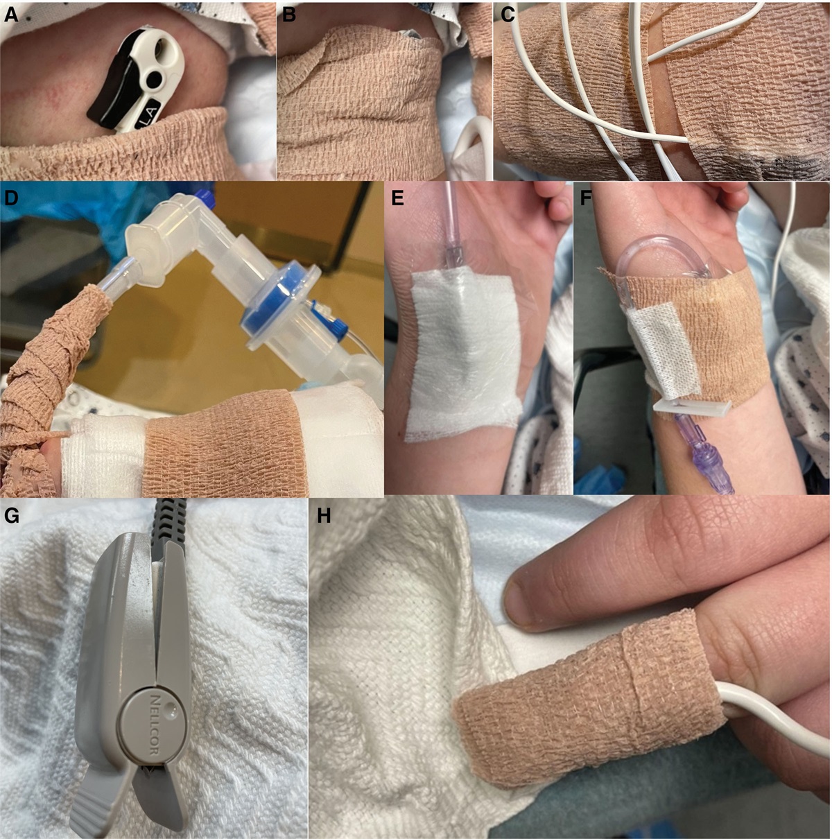
CASE REPORTA 43-year-old woman with past medical history of subacute cutaneous lupus erythematosus (SCLE), mild intermittent asthma, anxiety, and allergic rhinitis was scheduled for deep inferior epigastric perforator (DIEP) flap surgery after bilateral mastectomies for ductal carcinoma. Her outpatient medications included hydroxychloroquine, venlafaxine, anastrozole, and goserelin injections. During preoperative evaluation, it was noted that the patient had previously undergone 7 separate surgical procedures requiring anesthesia at multiple outside hospitals, and after each event, she suffered a rash of varying severity (Figures 1–3).
 Figure 1.:
 Painful rash present after previous surgery.
 Figure 2.:
 Painful rash present after previous surgery.
 Figure 3.:
 Painful rash present after previous surgery.
Ten years before the aforementioned DIEP flap procedure, the patient underwent an otherwise uneventful dilation and curettage. Two hours postoperatively, she had sudden onset of severe morbilliform rash beginning on her chest and neck, spreading throughout entire body, sparing only mucous membranes. The rash had a burning quality with some pruritis. The patient described it as “feeling like my skin was actually on fire.” The rash was associated with low-grade fevers and arthralgias, without evidence of hemodynamic changes, angioedema, or respiratory symptoms. Resolution occurred within 2 weeks without treatment. Subsequent investigation revealed the presence of antinuclear, Ro, and La antibodies, rheumatoid factor, and decreased complement factor components. Skin biopsy analysis was reported as potential “connective tissue disorder such as lupus.” She was diagnosed with SCLE.
Two years after this event, the patient underwent a rhinoplasty under general anesthesia and again suffered onset of a similar rash postoperatively. She was prescribed topical steroid cream and oral prednisone. The rash remained mild in intensity before complete resolution.
The patient was later diagnosed with breast cancer. Insertion of a port under monitored anesthesia care (MAC) resulted in onset of a mild rash, which was treated with a course of oral prednisone. After 5 months, she underwent a double mastectomy under general anesthesia with single-shot peripheral nerve blocks using liposomal bupivacaine. Again, approximately 2 hours after surgery, a “burning” morbilliform rash of moderate severity began. She was not given steroids due to concern for postsurgical healing. On postoperative day (POD) 1, she returned to surgery for evacuation of a breast hematoma; in the postanesthetic care unit (PACU), the rash became intensely painful. She was initially given topical steroid creams, but due to the diffuse progression of the rash, an oral prednisone course was eventually initiated. This rash resolved over 2 weeks.
The following year, she underwent a laparoscopic cholecystectomy under general anesthesia and again had onset of a rash 2 hours after surgery. She was treated with a course of oral prednisone, and the rash remained moderate in intensity. Two months later, she underwent an upper endoscopy under MAC, notably receiving only lidocaine, propofol, and fentanyl, and again suffered the rash.
Given the extensive history of recurrent postoperative rashes, the patient was referred to an allergy and immunology specialist for preoperative evaluation before DIEP flap. The specialist stated that previous rashes were likely medication reactions, with lidocaine, fentanyl, or propofol (the only medications given in all documented anesthetics) being the most likely triggers. However, the patient previously tolerated a labor epidural with bupivacaine and fentanyl, and multiple minor procedures under lidocaine, without resultant rashes. It was therefore deduced that propofol was the likely triggering agent.
After lengthy discussion of plans and risks with surgical and anesthesia teams, the patient agreed to DIEP flap procedure under general anesthesia with avoidance of propofol. At the instruction of the allergy and immunology specialist, 48 hours preoperatively the patient took oral prednisone 30 mg, cetirizine 20 mg, and famotidine 20 mg, and after inhalational induction, the patient was given diphenhydramine 50 mg intramuscular (IM) and hydrocortisone 100 mg intravenous (IV). Bilateral transversus abdominus plane and pectoralis nerve I and II blocks with bupivacaine 0.25% were placed after intubation. Intravenous opiate-based analgesics were reserved for postoperative care, if needed. The surgical case proceeded uneventfully for nearly 7 hours. Full-body skin examinations were performed hourly. Anesthetic emergence and extubation were unremarkable. The patient had no rash throughout PACU stay and did not require supplemental analgesics. She was discharged home on POD 1 and contacted daily for several days afterward. The patient never developed rash or other evidence of medication reaction, a first among her many postoperative courses.
One year later, the patient returned for bilateral reconstruction revision. This was performed under general anesthesia, again withholding propofol, but this time also without stress-dose steroids, cetirizine, or famotidine. Induction and maintenance of anesthesia was with sevoflurane. The patient received intravenous midazolam 2 mg, cefazolin 2 g, and dexamethasone 5 mg at the beginning of the case, and diphenhydramine 25 mg was given at emergence. The patient was monitored closely perioperatively for cutaneous changes; however, none developed.
DISCUSSIONPropofol is an anesthetic agent used for induction and maintenance of general anesthesia, as well as for sedation in perioperative and intensive care environments.1 The common side effect profile of propofol is well-described and includes hypotension, respiratory depression, injection site discomfort, myoclonus, and QT prolongation.2 Diligent review of the anesthetic records of this patient suggested propofol was likely associated with the patient’s postoperative rashes, a potential side effect of propofol that is infrequently described.3 By removing propofol from the anesthetic plans of both her DIEP flap and reconstruction revision, and observing no perioperative cutaneous changes for the first time in her lengthy surgical history, the case was strengthened that propofol had very likely been responsible for her 7 previous eruptions.
Antihistamines and stress-dose steroids were given during the DIEP flap, potentially preventing a rash that had an etiology other than PISCLE. However, antihistamines and low-dose steroids had also been given throughout some of her previous surgeries where postoperative rashes occurred, while she received no stress-dose steroids, cetirizine, or famotidine for her uneventful reconstruction revision. Propofol remains the only known variable present in all anesthetics after which a rash followed, and absent in both cases after which the patient remained asymptomatic.
There is little available literature describing similar dermatological reactions associated with propofol. Only a single case report from Yu et al3 was found during the literature review. This case report—which included images of a rash appearing remarkably similar to those taken of our patient’s rashes—reinforced the conclusion that propofol could be the agent responsible for the recurrent postoperative rashes and gave the authors the confidence to proceed with the elective procedure.
Yu et al proposed its skin manifestation was an acute exacerbation of systemic lupus erythematosus (SLE); it is unclear from the report if their patient had SLE at baseline. In our case, SCLE was diagnosed after multiple skin biopsies and seropositivity of several markers after her first postoperative rash. She did not meet the American College of Rheumatology’s diagnosis criteria for SLE.4,5 While patients with SCLE may meet criteria for SLE, the existence of both isolated idiopathic SCLE and DI-SCLE are well-documented in the literature.6,7 Key features that may distinguish DI-SCLE include widespread cutaneous lesions (particularly beyond sun-exposed areas), as well as the presence of lesions that are vasculitic-like, bullous, or erythema-multiforme-appearing.8
Cases of DI-SCLE have been frequently attributed to antihypertensives and antifungals, but such reactions to propofol remain largely undocumented.7 Most literature also documents DI-SCLE reactions with incubation periods of at least a week after drug exposure.9 However, these cases almost exclusively document reactions to oral medications, and the intravenous administration of propofol may account for its relatively fast onset of cutaneous symptoms.
Given the consistent presentation in both this case report and that from Yu et al, and the particular relevance of PISCLE to the field of anesthesiology, research into the true prevalence of similar reactions is needed. A valuable first step could include an online collaborative database where suspected cases can be submitted and analyzed. The pathophysiology of this condition will also require more investigation, but the increase in helper T-cell activity in both SLE patients and routine propofol exposure is a potential basis for exploration.3,10
Steroids anecdotally improved this patient’s clinical course during multiple cutaneous reactions, but the best management proved to be removal of propofol from her anesthetic plan. A heightened awareness of this uncommon reaction to propofol may produce more documented cases, facilitate quicker diagnoses with better management, and, ideally, help to identify other high-risk patients for whom it may be best to avoid propofol and prevent PISCLE.
DISCLOSURESName: Joseph A. Schoenfeldt, MD.
Contribution: This author helped make substantial contributions to the literature review, medical record review, writing of case report and discussion, research, and analysis.
Name: Michael A. Howard, MD.
Contribution: This author helped make substantial contributions to the literature review and editing, provided expert opinion, and helped with the research, analysis, and writing of this case report.
Name: Deeba Masood, MD.
Contribution: This author helped make substantial contributions to the literature review and editing, provided expert opinion, and helped with the research, analysis, and writing of this case report.
Name: Daniel S. Cormican, MD.
Contribution: This author helped make substantial contributions to the literature review and editing, provided expert opinion, and helped with the research, analysis, and writing of this case report.
This manuscript was handled by: Mark C. Phillips, MD.
REFERENCES 1. Folino TB, Muco E, Safadi AO, et al. Propofol. National Center for Biotechnology Information. Published May 8, 2022. Accessed July 6, 2022. Available at: https://pubmed.ncbi.nlm.nih.gov/28613634/. 2. DIPRIVAN (propofol) injectable emulsion, USP—Food and Drug Administration. Published April 2017. Accessed March 2, 2022. Available at: https://www.accessdata.fda.gov/drugsatfda_docs/label/2017/019627s066lbl.pdf. 3. Yu Y, Gui H, Xiong K, Fang X, He J. Progression of systemic lupus erythematosus associated with propofol administration: a case report. Eur J Hosp Pharm. 2018;25:225–227. 4. Tsokos GC. Systemic lupus erythematosus. N Engl J Med. 2011;365:2110–2121. 5. Ben-Menachem E. Systemic lupus erythematosus. Anesth Analg. 2010;111:665–676. 6. Tiao J, Feng R, Carr K, Okawa J, Werth VP. Using the American College of Rheumatology (ACR) and Systemic Lupus International Collaborating Clinics (SLICC) criteria to determine the diagnosis of systemic lupus erythematosus (SLE) in patients with subacute cutaneous lupus erythematosus (SCLE). J Am Acad Dermatol. 2016;74:862-869. 7. Lowe G, Henderson CL, Grau RH, Hansen CB, Sontheimer RD. A systematic review of drug-induced subacute cutaneous lupus erythematosus. Br J Dermatol. 2011;164:465–472. 8. Marzano AV, Lazzari R, Polloni I, Crosti C, Fabbri P, Cugno M. Drug-induced subacute cutaneous lupus erythematosus: evidence for differences from its idiopathic counterpart. Br J Dermatol. 2011;165:335–341. 9. Keyes E, Grinnell M, Vazquez T, Diaz D, Thomas P, Werth VP. Drug-induced subacute cutaneous lupus erythematosus in previously diagnosed systemic lupus erythematosus patients: a case series. JAAD Case Rep. 2021;12:18–21. 10. Pirttikangas CO, Perttilä J, Salo M, Vainio O, Liukko-Sipi S. Propofol infusion anaesthesia and immune response in minor surgery. Anaesthesia. 1994;49:13–16.
Comments (0)