
Remember me


Mucocutaneous manifestations of human immunodeficiency virus (HIV) are commonly seen in clinical practice; at least 90% of patients living with HIV (PLWHs) will develop some type of skin condition over the course of their lifetime. Skin is oftentimes the first organ affected by HIV and is a useful clinical marker of HIV disease progression (Basida et al., 2021). Mucocutaneous manifestations may include inflammatory conditions such as seborrheic dermatitis, pruritus, or psoriasis; viral, bacterial, and fungal infections; and HIV-associated malignancies like Kaposi sarcoma (KS; Chelidze et al., 2019). This article will review the most common dermatological conditions in PLWHs, as well as how they typically present in clinical practice, and discuss current evidence for treatment.
EPIDEMIOLOGYAlmost every PLWH will present with dermatological complaint at some point during their care. Compared with people living without HIV, PLWHs have a greater likelihood of skin eruptions because of dysregulation of the skin immune system or drug reactions (Ramos-e-Silva et al., 2020). A common skin condition that afflicts PLWHs is seborrheic dermatitis, which affects 30%–83% of patients (Adalsteinsson et al., 2020). Another common skin manifestation is methicillin-resistant Staphylococcus aureus (MRSA) infection; PLWHs have a 6- to 18-fold higher rate of infection with a recurrence rate of 27% within 6 months of treatment (Skiest et al., 2006). Mucocutaneous manifestations are typically more common as CD4 cell count drops, HIV RNA level increases, and immune function decreases. Studies found that pruritic papular rashes, psoriasis, and drug-induced reactions were more likely among PLWHs with CD4 < 200 (Esser & Sammet, 2021; Mirnezami et al., 2020). Some other conditions, such as varicella-zoster, may occur at any stages of HIV regardless of CD4 cell count; however, the manifestation may be more severe and extensive than compared with someone living without HIV (Chelidze et al., 2019).
PATHOGENESISThe skin's immune system maintains homeostasis of the body's internal operations by preventing and eliminating invasions from external pathogens, toxins, allergens, and neoplastic disorders (Barroca et al., 2014). This is accomplished through an immunological response process that involves the resident cells (kerantinocytes, lymphatic and capillary endothelial cells), antigen-presenting cells (Langerhans cells and dermal dendritic cells), and T-lymphocytes (Barroca et al., 2014). HIV infection disrupts this process, resulting in decreased numbers of Langerhans cells and dermal dendritic cells as well as CD4 T-lymphocytes, natural killer cells, macrophages, and monocytes. Any loss of immune cells allows the skin to become more vulnerable to opportunistic infections, inflammatory processes, and neoplastic disorders (Barroca et al., 2014). Clinically, the CD4 count is a frequently used parameter to determine HIV stage and aid in its management because HIV targets these cells. Although multiple variables can affect its count, a normal adult CD4 count, when taking in laboratory variations, may range from 500 to 1400 cells/μL (Sax, 2021). A CD4 count lower than 200 cells/μL is the clinical definition of acquired immunodeficiency syndrome and could aid in the clinical assessment of skin-related opportunistic infections (Sax, 2021).
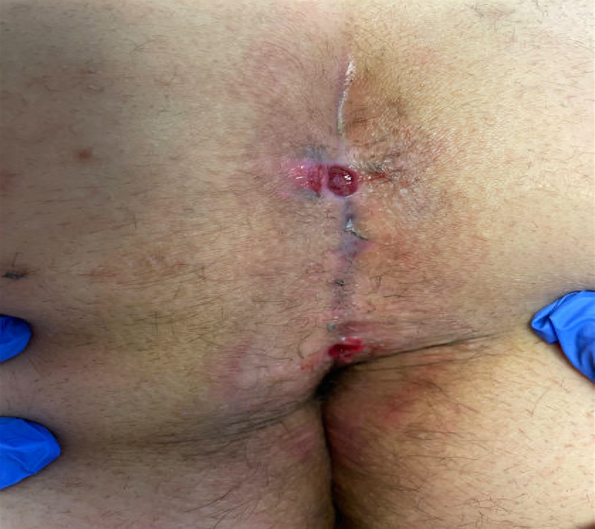
CUTANEOUS MANIFESTATIONS/CLINICAL FEATURES MRSA and Soft Tissue InfectionsAlthough there has been a decreased incidence of MRSA bacteremia with the introduction of antiretroviral therapy (ART), PLWHs are still at a higher risk of severe infection compared with those living without HIV, with lower CD4 count being one of the risk factors for MRSA (Lopez & Sanders, 2021; Shadyab & Crum-Cianflone, 2012). A review of literature found that the most common type of MRSA infections among PLWHs was soft tissue infections caused by the community strain USA300 (Shadyab & Crum-Cianflone, 2012). Localized skin and soft tissue infections are common and may present as abscesses, cellulitis, furuncles, folliculitis, ulcerations, and impetigo (Shadyab & Crum-Cianflone, 2012). Lesions are typically found on the lower and upper extremities, followed by the trunk, axillae, face, and neck with increased incidences found in the genital region (Shadyab & Crum-Cianflone, 2012). Risk factors to note in the clinical setting are poor immune status/lower CD4 count, high-risk sexual behaviors, illicit drug use, recent sexually transmitted infections, prior incarceration, and prior hospitalization, all of which require further interventions (Lopez & Sanders, 2021; Shadyab & Crum-Cianflone, 2012). Without treatment, localized skin infections in PLWHs may result in severe bacteremia, which may consequently lead to complications such as pneumonia, septic pulmonary emboli, osteomyelitis, meningitis, and endocarditis (Shadyab & Crum-Cianflone, 2012).
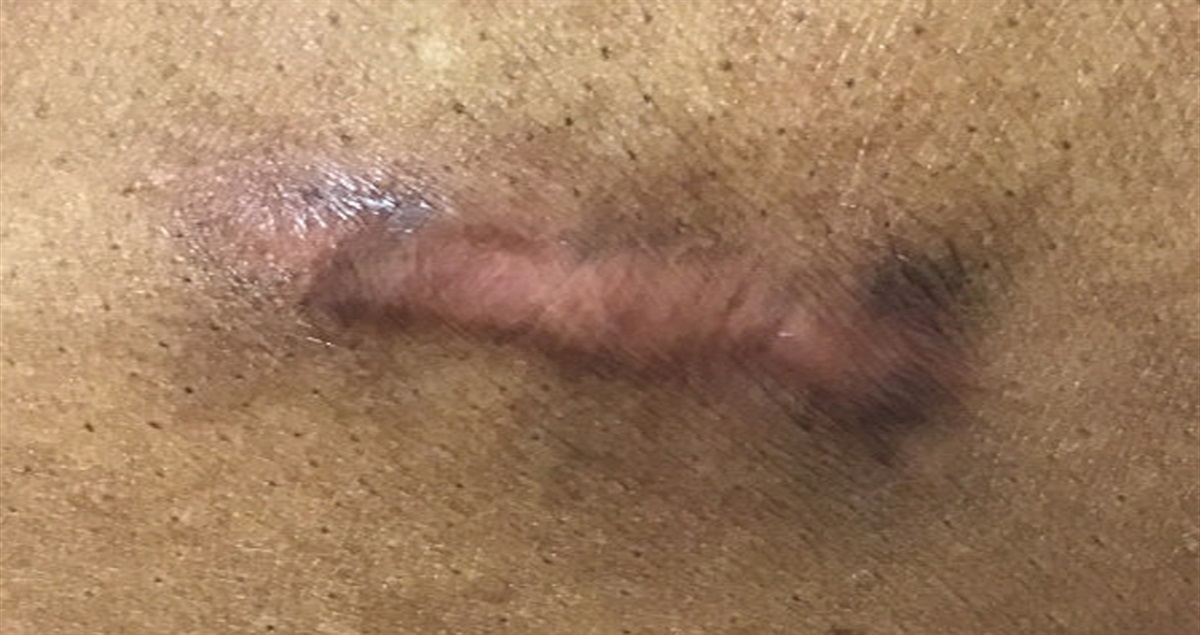
Bacillary AngiomatosisThis condition occurs because of infection by the bacteria Bartonella, with the species Bartonella henselae and Bartonella quintana affecting PLWHs more severely (Mosepele et al., 2012). Providers should be prompted to consider Bartonella infection if patients report being scratched by a cat, have lice, or may have experienced homelessness. Bacillary angiomatosis are typically seen in PLWHs with a CD4 cell count of <100 (Mosepele et al., 2012). Lesions may appear as reddish or purplish vascular nodules on the skin and are typically found in the upper extremities in clusters (Akram et al., 2022). PLWHs typically present with fever of unknown origin (≥38°C), osteomyelitis, and angioproliferative lesions affecting primarily the skin, liver, and spleen (Mosepele et al., 2012). Differential diagnoses to consider are KS, cherry angioma, or dermatofibroma given the similar presentation of these lesions to bacillary angiomatosis (Spach, 2022). It is important to note that bacillary angiomatosis, if misdiagnosed and treated with chemotherapy for presumed KS, can disseminate to the bone and viscera and become life-threatening (Forrestel et al., 2015). Histology should be performed to confirm diagnosis before beginning treatment (Forrestel et al., 2015).
Molluscum ContagiosumThis infectious viral condition manifests as discreet, skin-colored papules with a central umbilication measuring 2–5 mm (Chelidze et al., 2019; Spach, 2022). As HIV progresses and immune status weakens, these discreet lesions increase in numbers and become irregular in shape with loss of the umbilicated center; they may spread to the face and merge into larger lesions (“giant molluscum”) and may appear as disfiguring papular lesions similar to the morphology of warts (Chelidze et al., 2019).
Herpes ZosterThe highest risk for reactivation of varicella-zoster virus among PLWHs is in those with CD4 cell counts of less than 200 cells/mm3 and in those who started ART within the first 4 months because of immune reconstitution inflammatory syndrome (IRIS; Chelidze et al., 2019). Physical manifestations are usually preceded by prodrome of tingling/numbness. Lesions appear in clusters of vesicles on an erythematous base distributed on dermatomes but may also appear as chronic, ulcerative, bullous, or verrucous lesions (Chelidze et al., 2019). Once healed, PLWHs are at a higher risk of developing postherpetic neuralgia compared with those without HIV (Forbes et al., 2016). Those with severely immunocompromised status, including PLWHs, have a higher likelihood of developing disseminated infection to the eyes, central nervous system, or visceral organs (Spach, 2021).
Seborrheic DermatitisSkin biopsies showed a different histology in seborrheic lesions among PLWHs and those in the general population (Cedeno-Laurent et al., 2011; Mirnezami et al., 2020). In the general population, seborrheic lesions may present initially on the scalp, resulting in dandruff, or on the back or shoulder; clinical findings may show greasy scales above discreet, well-approximated erythematous patches. For PLWHs, erythema is more widespread, lesions are more extensive and may be found in areas beyond the face, scalp, shoulder, and back; and a biopsy of lesions shows proteins otherwise not seen in those without HIV (Adalsteinsson et al., 2020; Cedeno-Laurent et al., 2011).
Scabies is a parasitic condition that is transmitted by skin-to-skin contact and results in intense pruritus for the patient. It may appear initially as small erythematous papules and turn to vesicular lesions (Chosidow, 2006). The hallmark of scabies is thin, short, wavy burrows in the spaces between fingers, wrist flexors, axillae, feet, genitalia in men, areola in women, and inner thighs (Thomas et al., 2020). Crusted scabies, on the other hand, differ from common scabies in that they are often seen in PLWHs with severe immunodeficiency and have a hyperkeratotic scaly appearance on the buttocks (Spach, 2021; Tolkachjov et al., 2018).
Herpes Simplex VirusPLWHs with herpes simplex virus (HSV) frequently present with atypical lesions that are extensive, painful, and prolonged, especially among those with severe immunosuppression (Chelidze et al., 2019). Lesions may appear on the lips or mouth with HSV-1 and around the genitalia region if patients have HSV-2, although location is not definitive of HSV type (Burke & Lopez, 2017). HSV typically presents initially with prodromal sensory symptoms, progressing to physical manifestation of papular and then vesicular lesions, followed by the crusting stage (Burke & Lopez, 2017). Clinicians should be aware that chronic, painful, nonhealing ulcers are hallmarks of severe immunodeficiency (CD4 cell count below 100 cells/mm3) and can occur anywhere on the body aside from the lips, mouth, or genitals (Chelidze et al., 2019). One study found that although the frequency of lesions did not change ART initiation, long-term ART use reduced both HSV shedding and recurrence of lesions (Ford et al., 2018).
SyphilisThe cutaneous manifestations of syphilis are similar among PLWHs and those without HIV (Chelidze et al., 2019). In PLWHs, primary syphilis may manifest as many large, slow-healing chancres compared with those without HIV and may be atypical or painful (Chelidze et al., 2019; Workowski et al., 2021). Secondary syphilis on examination may reveal maculopapular lesions on the trunks and extremities with palm and sole involvement, as well as uncommonly seen cutaneous manifestations such as annular plaques with hypopigmented centers, or necrotic, crusty, and scaly plaques (Chelidze et al., 2019). Clinicians should recognize that PLWHs may present with both primary and secondary syphilis and may quickly progress to tertiary syphilis (Chelidze et al., 2019). Clinicians should also conduct a thorough ocular examination as ocular syphilis and associated neurosyphilis have been rising in recent years (Chelidze et al., 2019).
Human PapillomavirusCompared with those living without HIV, PLWHs are more likely to present with human papillomavirus (HPV) infection (Chelidze et al., 2019). The etiology has been suggested to relate to the dysregulation of p27 expression and proliferation of cutaneous cells (Chelidze et al., 2019). Warts may appear in genital or sun-exposed areas of the body and present with more severity and with a higher frequency than those living without HIV (Coates & Leslie, 2019). In women, HPV infection and progression of HPV-associated cervical lesions are highly associated with an immunodeficient status (G. Liu et al., 2018).
PsoriasisAlthough the prevalence of psoriasis among PLWHs and those in the general population is similar, lesions among PLWHs are typically more severe, destructive, and resistant to treatment (Ceccarelli et al., 2019; Cedeno-Laurent et al., 2011). Psoriasis lesions among PLWHs tend to present as extensive lesions of guttate, inverse, and erythrodermic subtypes as well as those affecting the extremities, fingers, or ears (Ceccarelli et al., 2019; Cedeno-Laurent et al., 2011; Spach, 2021). Clinicians should also assess for psoriatic arthritis among those who are severely immunocompromised, including PLWHs, as it is common in this population (Cedeno-Laurent et al., 2011). Psoriatic arthritis in PLWHs may present as joint pain, stiffness, and effusion in the finger joints and spine (Spach, 2021).
Eosinophilic FolliculitisThe condition is typically seen in PLWHs and more often in those with advanced disease (Cedeno-Laurent et al., 2011). Eosinophilic folliculitis usually presents as a papular rash on an erythematous base with scattered vesicular or pustular lesions on the face, neck, chest, and back, primarily above the nipple line (Afonso et al., 2012; Cedeno-Laurent et al., 2011). Laboratory findings reveal elevated serum immunoglobulin E levels, eosinophilia, and peripheral leukocytosis (Cedeno-Laurent et al., 2011).
Prurigo NodularisThis is a cutaneous manifestation that occurs in both PLWHs and those without HIV. The lesions commonly present as nodular or papular hyperkeratotic lesions approximately 3–5 mm in size with associated crusting in a symmetrical distribution (Garg & Sanke, 2017; Matthews & Cockerell, 1998). Lesions are typically seen on the trunk and extremities without face or scalp involvement (Whang et al., 2019). The patient will typically complain of generalized, intense pruritus that causes scratching to relieve symptoms (Whang et al., 2019). It is important for clinicians to distinguish this condition from other similar skin manifestations such as eosinophilic folliculitis given similar presentation to the affected areas.
Drug-Induced ReactionsPLWHs are at a higher risk of cutaneous reactions to medications compared with the general population (Cedeno-Laurent et al., 2011; Chelidze et al., 2019). These reactions may include mild and more common cases such as morbilliform exanthematous reactions or more severe cases such as Stevens–Johnson syndrome and toxic epidermal necrolysis (Cedeno-Laurent et al., 2011). Mucocutaneous presentations may include pruritic maculopapular eruptions, generalized urticaria or angioedema, acute generalized erythematous pustulosis, drug rashes with eosinophilia and systemic symptoms, and xerosis. Clinicians should be aware that certain ART medications, including protease inhibitors, efavirenz, nevirapine, and abacavir, are more likely to cause reactions (Cedeno-Laurent et al., 2011). PLWHs with a CD4 cell count below 200 cells/mm3 are typically placed on sulfamethoxazole/trimethoprim for opportunistic infection prophylaxis, which may also induce a cutaneous reaction (typically a maculopapular rash with or without a fever) and require further evaluation (Cedeno-Laurent et al., 2011; Giles et al., 2019). Compared with the 3%–8% rate of sulfa hypersensitivity in the general population, a study found that 27% of PLWHs developed drug-induced reactions when taking sulfamethoxazole/trimethoprim for the treatment of pneumocystis pneumonia (Giles et al., 2019). It is important to perform a thorough history to differentiate between drug-induced reactions from other causes, such as IRIS. IRIS is a hyperinflammatory response to latent infections after patients initiate ART and their CD4 count improves; these infections may include pneumocystis pneumonia, cryptococcal meningitis, or Hepatitis B or C (Thapa & Shrestha, 2022).
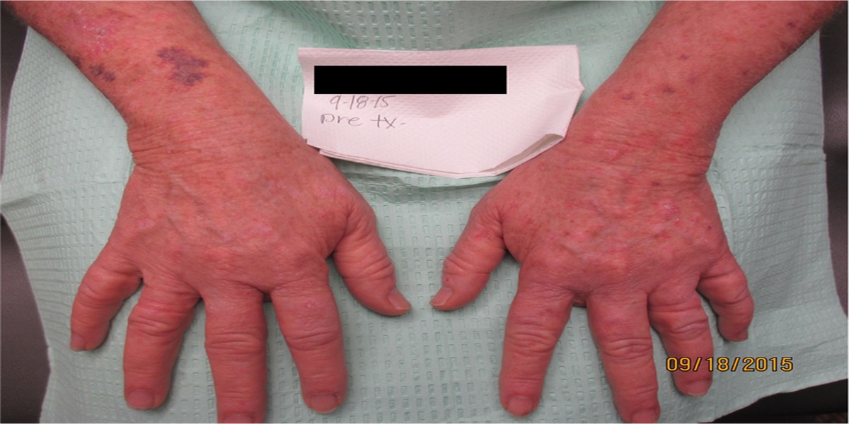
Kaposi SarcomaKS is a vascular malignancy caused by the oncovirus human herpes virus 8 and primarily affects PLWHs who are severely immunosuppressed and may also present in older men and transplant patients (Cesarman et al., 2019; Coates & Leslie, 2019). Since the initiation of ART in the 1990s, there has been a reduction of incidence in KS among PLWHs in the United States. However, the risk of KS remains extremely high among PLWHs compared with those who do not have HIV (Coates & Leslie, 2019). KS can present as violaceous macular, papular, or plaque lesions. KS affects the skin but can also involve other organs such as the lungs and gastrointestinal tract (Ramos-e-Silva et al., 2020). Recent research suggested that cumulative viral load level may trigger early development of KS independent of CD4 cell count; therefore, clinicians should be concerned of KS regardless of CD4 cell count and perform a thorough physical examination at every patient encounter (Dubrow et al., 2017). For confirmatory diagnosis of KS, a biopsy of the lesion should be sent for pathology (Dubrow et al., 2017).
DIFFERENTIAL DIAGNOSES OF SKIN MANIFESTATIONSGiven the numerous expressions of skin lesions that present in clinical practice among PLWHs, it may be appropriate to break down the differential diagnoses by categorizing skin manifestations based on their morphological appearances. Given that PLWHs have higher incidence of bacterial infections, providers should consider soft tissue and Staphylococcus aureus infections if lesions appear to be purulent or are abscess forming (Lopez & Sanders, 2021). Macular lesions may suggest cutaneous drug eruption, acute HIV infection, or primary and secondary syphilis infection (Spach, 2021). Papular lesions may include differential diagnoses of molluscum contagiosum, eosinophilic folliculitis, warts, and scabies (Spach, 2021). Clinicians should be prompted to consider KS, bacillary angiomatosis, or prurigo nodularis when encountering nodular lesions (Spach, 2021). Scaling of the skin may indicate seborrheic dermatitis or psoriasis as well as the final stage of HSV infection (Spach, 2021). Vesicular lesions may indicate HSV or varicella-zoster virus (Spach, 2021). Providers should further narrow down their differential diagnoses by pattern of lesions, onset, location, symptomology, patient's immune status, and laboratory testing or biopsy, as appropriate.
It is important to recognize that certain mucocutaneous lesions present differently in PLWHs versus the general population. For example, the subtypes of psoriasis that are commonly noted in PLWHs are inverse, guttate, erythrodermic, and acral lesions with associated symptoms of joint pain, versus the classical presentation of plaque-like lesions (Cedeno-Laurent et al., 2011). HSV lesions may be extensive, ulcerative, painful, and refractory to treatment, especially if patients are immunocompromised and/or not on ART (Chelidze et al., 2019). Understanding the unique presentations of these mucocutaneous conditions may aid the clinicians in the development of an appropriate care plan (e.g., initiating ART, supportive management).
Recognizing the patient's immune status is also critical in understanding how mucocutaneous lesions may present and where. For example, psoriasis is a T-lymphocyte-mediated disease that improves or worsens based on immune status; therefore, initiation of ART and return of CD4 cells among PLWHs with psoriasis will show most significant improvements in psoriasis eruptions (Menon et al., 2010).
Once a diagnosis is established, clinicians may continue with treatment of the skin condition per the most recent, evidence-based guidelines.
TREATMENT OF SKIN MANIFESTATIONSTable 1 below summarizes the treatments for common mucocutaneous conditions described in this article and may be utilized as a quick guide in the clinical setting. CD4 count should be used as a guide but not a definitive approach when diagnosing a dermatological condition, as CD4 count may fluctuate depending on other comorbidities or conditions and not a true marker of HIV progression (Sax, 2021). For many dermatological conditions related to HIV, initiation and continuation of ART should be considered as the first line of treatment in conjunction with other measures as many skin conditions are associated with poor prognosis and a faster progression to acquired immunodeficiency syndrome (Lopez & Sanders, 2021).
TABLE 1 - Summary of Treatments for Common Mucocutaneous Conditions Cutaneous Manifestation Immune Status (CD4 Cells/mm 3) Treatment Clinical Practice Guidelines or Evidence-Based Practice Bacterial MRSA and soft tissue infections Any CD4 count In the outpatient setting: incision and drainage of abscess and addition of antibiotics if: Patients have an extensive disease with evidence of cellulitis Systemic illness Comorbidities and immunosuppression Patients are older adults Lack of response to incision and drainage or area difficult to drain For purulent cellulitis, cover for community-acquired MRSA with any of the following antibiotics, and duration may last 5–10 days: Clindamycin 300–450 mg PO TIDTreatments listed in this table do not address treatment for pregnant women or children; refer to the clinical guidelines for specific treatment recommendations for these populations. In addition, this table is not all-encompassing of treatments for various skin manifestations of HIV; therefore, providers should refer to clinical guidelines for full treatment information. MRSA = methicillin-resistant Staphylococcus aureus; ART = antiretroviral therapy; IRIS = immune reconstitution inflammatory syndrome; HIV = human immunodeficiency virus; PLWHs = patients living with HIV; HSV = herpes simplex virus; HPV = human papillomavirus.
aSubclinical shedding may still occur; symptoms may worsen early after initiation of ART.
bTreatment courses may need to be extended.
cSymptoms may worsen early after initiation of ART in the setting of IRIS.
There are over 50 dermatological conditions found to be associated with HIV infection (Cedeno-Laurent et al., 2011). The mucocutaneous clinical presentation for PLWHs is diverse and may vastly vary from the general population. Nurses and nurse practitioners providing care to this population should be well versed in the most common types of dermatological conditions with which patients may present. Furthermore, providers should be aware of how a patient's immune status plays a role in the mucocutaneous manifestations of various skin conditions as well as course of treatment. It is imperative that patients who are newly diagnosed with HIV are immediately started on ART, as viral suppression and immune function recovery are paramount to alleviating many skin conditions discussed in this article.

 REFERENCES
Adalsteinsson J. A., Kaushik S., Muzumdar S., Guttman-Yassky E., Ungar J. (2020). An update on the microbiology, immunology and genetics of seborrheic dermatitis. Experimental Dermatology, 29(5), 481–489. 10.1111/exd.14091
Afonso J. P. J. M., Tomimori J., Michalany N. S., Nonogaki S., Porro A. M. (2012). Pruritic papular eruption and eosinophilic folliculitis associated with human immunodeficiency virus (HIV) infection: A histopathological and immunohistochemical comparative study. Journal of the American Academy of Dermatology, 67(2), 269–275. 10.1016/j.jaad.2011.11.923
Akram S. M., Anwar M. Y., Thandra K. C., Rawla P. (2022). Bacillary angiomatosis. StatPearls Publishing. https://www.ncbi.nlm.nih.gov/books/NBK448092/
Barroca P., Calado M., Azevedo-Pereira J. M. (2014). HIV/dendritic cell interaction: Consequences in the pathogenesis of HIV infection. AIDS Reviews, 16(4), 223–235.
Basida S. D., Basida B., Zalavadiya N., Trivedi A. P. (2021). Dermatological opportunistic infections in HIV seropositive patients: An observational study. Cureus, 13(8), e16852. 10.7759/cureus.16852
Borda L. J., Perper M., Keri J. E. (2019). Treatment of seborrheic dermatitis: A comprehensive review. Journal of Dermatological Treatment, 30(2), 158–169. 10.1080/09546634.2018.1473554
Burke V. E., Lopez F. A. (2017). Approach to skin and soft tissue infections in non-HIV immunocompromised hosts. Current Opinion in Infectious Diseases, 30(4), 354–363. 10.1097/QCO.0000000000000378
Ceccarelli M., Venanzi Rullo E., Vaccaro M., Facciolà A., d'Aleo F., Paolucci I. A., Cannavo S. P., Cacopardo B., Pinzone M. R., Pellicanò G. F., Condorelli F., Nunnari G., Guarneri C. (2019). HIV-associated psoriasis: Epidemiology, pathogenesis, and management. Dermatologic Therapy, 32(2), e12806. 10.1111/dth.12806
Cedeno-Laurent F., Gómez-Flores M., Mendez N., Ancer-Rodríguez J., Bryant J. L., Gaspari A. A., Trujillo J. R. (2011). New insights into HIV-1-primary skin disorders. Journal of the International AIDS Society, 14, 5. 10.1186/1758-2652-14-5
Cesarman E., Damania B., Krown S. E., Martin J., Bower M., Whitby D. (2019). Kaposi sarcoma. Nature reviews. Disease Primers, 5(1), 9. https://doi.org/10.1038/s41572-019-0060-9
Chelidze K., Thomas C., Chang A. Y., Freeman E. E. (2019). HIV-related skin disease in the era of antiretroviral therapy: Recognition and management. American Journal of Clinical Dermatology, 20(3), 423–442. 10.1007/s40257-019-00422-0
Chosidow O. (2006). Scabies. New England Journal of Medicine, 354(16), 1718–1727. 10.1056/NEJMcp052784
Coates S. J., Leslie K. S. (2019). What's new in HIV dermatology? F1000Res, 8, F1000. 10.12688/f1000research.16182.1
Dubrow R., Qin L., Lin H., Hernández-Ramírez R. U., Neugebauer R. S., Leyden W., Alhoff K. N., Achenbach C. J., Hessol N. A., Modur S. P., D'Souza G., Bosch R. J., Grover S., Horberg M. A., Kitahata M. M., Mayor A. M., Novak R. M., Rabkin C. S., Sterling T. R., Silverberg M. J.; North American AIDS Cohort Collaboration on Research and Design of the International Epidemiologic Databases to Evaluate AIDS. (2017). Associations of CD4+ T-cell count, HIV-1 RNA viral load, and antiretroviral therapy with Kaposi sarcoma risk among HIV-infected persons in the United States and Canada. Journal of Acquired Immune Deficiency Syndromes, 75(4), 382–390. 10.1097/QAI.0000000000001394
Esser S., Sammet S. (2021). HIV medicine for dermatologists and venereologists. Journal of the German Society of Dermatology, 19(1), 82–96. 10.1111/ddg.14373
Forbes H. J., Bhaskaran K., Thomas S. L., Smeeth L., Clayton T., Mansfield K., Minassian C., Langan S. M. (2016). Quantification of risk factors for postherpetic neuralgia in herpes zoster patients: A cohort study. Neurology, 87(1), 94–102. 10.1212/WNL.0000000000002808
Ford E. S., Magaret A. S., Spak C. W., Selke S., Kuntz S., Corey L., Wald A. (2018). Increase in HSV shedding at initiation of antiretroviral therapy and decrease in shedding over time on antiretroviral therapy in HIV and HSV-2 infected persons. AIDS, 32(17), 2525–2531. 10.1097/qad.0000000000002002
Forrestel A. K., Naujokas A., Martin J. N., Maurer T. A., McCalmont T. H., Laker-Opwonya M. O., Mulyowa G., Busakhala N., Amerson E. H. (2015). Bacillary angiomatosis masquerading as Kaposi's sarcoma in East Africa. Journal of the International Association of Providers of AIDS Care, 14(1), 21–25. 10.1177/2325957414521497
Garg T., Sanke S. (2017). Inflammatory dermatoses in human immunodeficiency virus. Indian Journal of Sexually Transmitted Diseases and AIDS, 38(2), 113–120. 10.4103/ijstd.IJSTD_22_17
Giles A., Foushee J., Lantz E., Gumina G. (2019). Sulfonamide allergies. Pharmacy (Basel), 7(3), 132. 10.3390/pharmacy7030132
Gupta A. K., Bluhm R. (2004). Seborrheic dermatitis. Journal of the European Academy of Dermatology and Venereology, 18(1), 13–20. https://doi.org/10.1111/j.1468-3083.2004.00693.x
Liu C., Bayer A., Cosgrove S. E., Daum R. S., Fridkin S. K., Gorwitz R. J., Kaplan S. L., Karchmer A. W., Levine D. P., Murray B. E., Rybak M. J., Telan D. A., Chambers H. F.; Infectious Diseases Society of America. (2011). Clinical practice guidelines by the Infectious Diseases Society of America for the treatment of methicillin-resistant Staphylococcus aureus infections in adults and children. Clinical Infectious Diseases, 52(3), e18–e55. 10.1093/cid/ciq146
Liu G., Sharma M., Tan N., Barnabas R. V. (2018). HIV-positive women have higher risk of human papilloma virus infection, precancerous lesions, and cervical cancer. AIDS, 32(6), 795–808. 10.1097/QAD.0000000000001765
REFERENCES
Adalsteinsson J. A., Kaushik S., Muzumdar S., Guttman-Yassky E., Ungar J. (2020). An update on the microbiology, immunology and genetics of seborrheic dermatitis. Experimental Dermatology, 29(5), 481–489. 10.1111/exd.14091
Afonso J. P. J. M., Tomimori J., Michalany N. S., Nonogaki S., Porro A. M. (2012). Pruritic papular eruption and eosinophilic folliculitis associated with human immunodeficiency virus (HIV) infection: A histopathological and immunohistochemical comparative study. Journal of the American Academy of Dermatology, 67(2), 269–275. 10.1016/j.jaad.2011.11.923
Akram S. M., Anwar M. Y., Thandra K. C., Rawla P. (2022). Bacillary angiomatosis. StatPearls Publishing. https://www.ncbi.nlm.nih.gov/books/NBK448092/
Barroca P., Calado M., Azevedo-Pereira J. M. (2014). HIV/dendritic cell interaction: Consequences in the pathogenesis of HIV infection. AIDS Reviews, 16(4), 223–235.
Basida S. D., Basida B., Zalavadiya N., Trivedi A. P. (2021). Dermatological opportunistic infections in HIV seropositive patients: An observational study. Cureus, 13(8), e16852. 10.7759/cureus.16852
Borda L. J., Perper M., Keri J. E. (2019). Treatment of seborrheic dermatitis: A comprehensive review. Journal of Dermatological Treatment, 30(2), 158–169. 10.1080/09546634.2018.1473554
Burke V. E., Lopez F. A. (2017). Approach to skin and soft tissue infections in non-HIV immunocompromised hosts. Current Opinion in Infectious Diseases, 30(4), 354–363. 10.1097/QCO.0000000000000378
Ceccarelli M., Venanzi Rullo E., Vaccaro M., Facciolà A., d'Aleo F., Paolucci I. A., Cannavo S. P., Cacopardo B., Pinzone M. R., Pellicanò G. F., Condorelli F., Nunnari G., Guarneri C. (2019). HIV-associated psoriasis: Epidemiology, pathogenesis, and management. Dermatologic Therapy, 32(2), e12806. 10.1111/dth.12806
Cedeno-Laurent F., Gómez-Flores M., Mendez N., Ancer-Rodríguez J., Bryant J. L., Gaspari A. A., Trujillo J. R. (2011). New insights into HIV-1-primary skin disorders. Journal of the International AIDS Society, 14, 5. 10.1186/1758-2652-14-5
Cesarman E., Damania B., Krown S. E., Martin J., Bower M., Whitby D. (2019). Kaposi sarcoma. Nature reviews. Disease Primers, 5(1), 9. https://doi.org/10.1038/s41572-019-0060-9
Chelidze K., Thomas C., Chang A. Y., Freeman E. E. (2019). HIV-related skin disease in the era of antiretroviral therapy: Recognition and management. American Journal of Clinical Dermatology, 20(3), 423–442. 10.1007/s40257-019-00422-0
Chosidow O. (2006). Scabies. New England Journal of Medicine, 354(16), 1718–1727. 10.1056/NEJMcp052784
Coates S. J., Leslie K. S. (2019). What's new in HIV dermatology? F1000Res, 8, F1000. 10.12688/f1000research.16182.1
Dubrow R., Qin L., Lin H., Hernández-Ramírez R. U., Neugebauer R. S., Leyden W., Alhoff K. N., Achenbach C. J., Hessol N. A., Modur S. P., D'Souza G., Bosch R. J., Grover S., Horberg M. A., Kitahata M. M., Mayor A. M., Novak R. M., Rabkin C. S., Sterling T. R., Silverberg M. J.; North American AIDS Cohort Collaboration on Research and Design of the International Epidemiologic Databases to Evaluate AIDS. (2017). Associations of CD4+ T-cell count, HIV-1 RNA viral load, and antiretroviral therapy with Kaposi sarcoma risk among HIV-infected persons in the United States and Canada. Journal of Acquired Immune Deficiency Syndromes, 75(4), 382–390. 10.1097/QAI.0000000000001394
Esser S., Sammet S. (2021). HIV medicine for dermatologists and venereologists. Journal of the German Society of Dermatology, 19(1), 82–96. 10.1111/ddg.14373
Forbes H. J., Bhaskaran K., Thomas S. L., Smeeth L., Clayton T., Mansfield K., Minassian C., Langan S. M. (2016). Quantification of risk factors for postherpetic neuralgia in herpes zoster patients: A cohort study. Neurology, 87(1), 94–102. 10.1212/WNL.0000000000002808
Ford E. S., Magaret A. S., Spak C. W., Selke S., Kuntz S., Corey L., Wald A. (2018). Increase in HSV shedding at initiation of antiretroviral therapy and decrease in shedding over time on antiretroviral therapy in HIV and HSV-2 infected persons. AIDS, 32(17), 2525–2531. 10.1097/qad.0000000000002002
Forrestel A. K., Naujokas A., Martin J. N., Maurer T. A., McCalmont T. H., Laker-Opwonya M. O., Mulyowa G., Busakhala N., Amerson E. H. (2015). Bacillary angiomatosis masquerading as Kaposi's sarcoma in East Africa. Journal of the International Association of Providers of AIDS Care, 14(1), 21–25. 10.1177/2325957414521497
Garg T., Sanke S. (2017). Inflammatory dermatoses in human immunodeficiency virus. Indian Journal of Sexually Transmitted Diseases and AIDS, 38(2), 113–120. 10.4103/ijstd.IJSTD_22_17
Giles A., Foushee J., Lantz E., Gumina G. (2019). Sulfonamide allergies. Pharmacy (Basel), 7(3), 132. 10.3390/pharmacy7030132
Gupta A. K., Bluhm R. (2004). Seborrheic dermatitis. Journal of the European Academy of Dermatology and Venereology, 18(1), 13–20. https://doi.org/10.1111/j.1468-3083.2004.00693.x
Liu C., Bayer A., Cosgrove S. E., Daum R. S., Fridkin S. K., Gorwitz R. J., Kaplan S. L., Karchmer A. W., Levine D. P., Murray B. E., Rybak M. J., Telan D. A., Chambers H. F.; Infectious Diseases Society of America. (2011). Clinical practice guidelines by the Infectious Diseases Society of America for the treatment of methicillin-resistant Staphylococcus aureus infections in adults and children. Clinical Infectious Diseases, 52(3), e18–e55. 10.1093/cid/ciq146
Liu G., Sharma M., Tan N., Barnabas R. V. (2018). HIV-positive women have higher risk of human papilloma virus infection, precancerous lesions, and cervical cancer. AIDS, 32(6), 795–808. 10.1097/QAD.0000000000001765
Comments (0)