
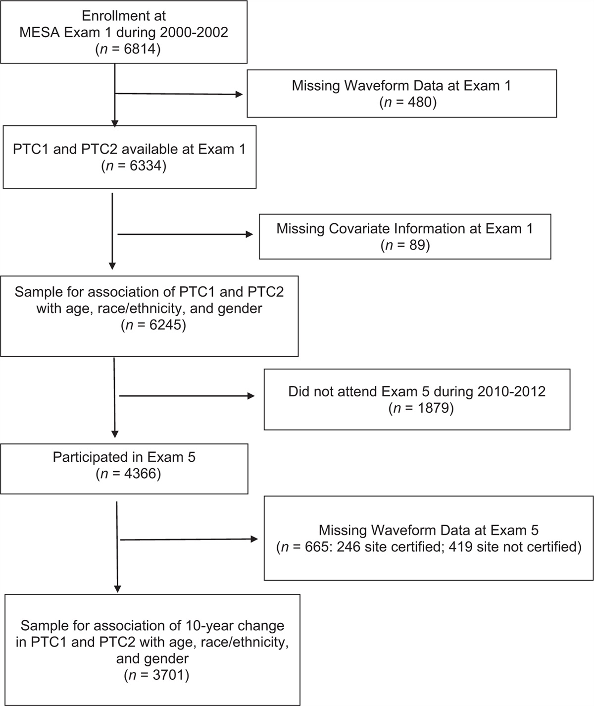
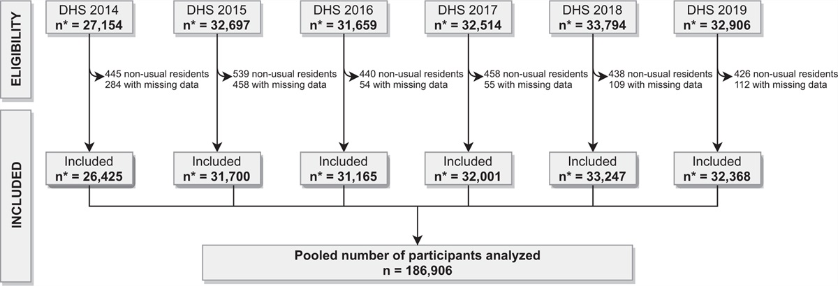
Remember me


The arterial baroreceptor reflex is a crucial blood pressure (BP) regulatory mechanism that maintains BP within a relatively narrow range of fluctuations [1,2]. When the baroreceptors within the carotid sinuses and aortic arch are loaded, the reflex pathway inhibits sympathetic outflow to the heart and vasculature and augments parasympathetic drive to the heart. This synchronal action restores baseline BP levels by promoting arterial vasodilation and reductions in cardiac output [1,2]. Impaired baroreflex function and sympathetic hyperactivity have been shown to be part of the pathophysiology of chronic hypertension [3–5], and now with the rapid advancement in technology for the precision detection and modulation of electrical signaling patterns within the nervous system, new interventional strategies such as baroreflex activation therapy (BAT) have emerged. Indeed, this novel treatment approach has been successfully applied to the baroreflex arc in humans to treat resistant hypertensive patients [6]. This surgical intervention, which delivers controlled electrical impulses to the carotid baroreceptors, has been quite promising, with clinical studies demonstrating meaningful reductions in BP and overall cardiovascular risk in the hypertensive population [7–9]. Despite these advances, our knowledge of the anatomical and functional aspects of the arterial baroreflex and how these elements impact cardiovascular disease progression in hypertension is still limited.
Electrical activation of the baroreceptor afferent fibers within the aortic depressor nerve (ADN) in rats represents a reliable method for exploring physiological and pathophysiological mechanisms pertaining to baroreflex-driven cardiovascular regulation [10,11]. In particular, direct electrical stimulation of the baroreceptor afferents in spontaneously hypertensive rats (SHR) carries a massive potential to functionally assess the baroreflex circuit in relation to hypertension, identify novel targets for BP modulation and discover new methods for enhancing and improving current BAT technology. Indeed, using the SHR model, we have recently identified the ‘aortic’ baroreceptor afferents as a viable alternative neuromodulation target to ‘carotid’ baroreceptor stimulation in lowering mean arterial pressure (MAP) [12]. We have further showed that ‘low intensity’ stimulation is an effective alternate way for neuromodulation of the aortic baroreceptor afferents, enabling low-energy consumption to induce a clinically relevant reduction in MAP levels in the SHRs [12]. Those recent findings, however, were primarily based on documenting responses to unilateral activation of the ‘left’ aortic baroreceptor afferents, without taking into account whether similar outcomes can be generated when the baroafferent fibers within the contralateral side are activated or when the afferent fibers on both sides are recruited simultaneously. The number of reports supporting anatomical and functional differences in the left versus right baroreflex pathways is quite limited, and this scarcity of information is even more pronounced under pathological conditions, such as hypertension. In normotensive Sprague–Dawley rats, we were able to demonstrate laterization in aortic baroreflex control of circulatory hemodynamics, whereby activation of the left ADN evoked greater reductions in MAP relative to right-sided stimulation, and bilateral stimulation of the ADN was as effective as the left-sided stimulation in promoting reflex hypotension [13].
Anatomically, the left baroreceptor afferents in rats innervate the aortic arch at the junction region of the left common carotid and left subclavian arteries [14]. In contrast, the right afferent bundles, which enter the junction region of the right common carotid artery and right subclavian artery, project onto the lower right common carotid artery and are consequently placed further away from the heart slightly above the aortic arch level [14]. It has also been demonstrated that the left ADN tends to carry a greater number of axons compared with the right ADN [14]. These anatomical data tally quite well with the above functional data, suggesting that left-sided dominance in the expression of ‘aortic’ baroreflex function is likely contributed to by the larger number of axons in the left ADN and closer positioning of afferent fibers to the heart relative to those of the right side. In the SHRs, the hypertensive state is associated with smaller ADN myelinated (A-type) and unmyelinated (C-type) axonal diameter, thinner myeline sheath around myelinated axons, and smaller number of unmyelinated fibers [15–17], all are elements which are thought to underlie altered baroreflex function in the SHRs. In view of the above functional and anatomical evidence, it remains unknown if laterality in the expression of ‘aortic’ baroreflex function is lost, altered, or maintained in the SHR model. This study, therefore, aimed to investigate left–right asymmetry in the processing of ‘aortic’ baroreceptor afferent input in the SHRs and how left-sided, right-sided, and bilateral activation of the ADN would impact reflex control of circulatory functions.
MATERIALS AND METHODS AnimalsAll experimental procedures adhered to the National Institutes of Health Guide for the Care and Use of Laboratory Animals prepared by the National Academy of Sciences and published by the National Institutes of Health. The protocols were reviewed and approved by the Animal Care and Use Committee of Case Western Reserve University.
Adult male SHRs weighing 320–390 g (total n = 9) were sourced from Envigo, USA. All animals were maintained in a controlled environment under a 12 h light/dark cycle and provided with rat chow and water ad libitum.
Surgical proceduresRats were anesthetized with an intraperitoneal injection of 50 mg/kg of sodium pentobarbital (Diamondback, Arizona, USA), and anesthesia was maintained using a continuous infusion (Harvard Apparatus Ltd., Holliston, Massachusetts, USA) of the anesthetic infused into the right femoral vein. Maintenance anesthesia was calculated at a dose of 10 mg/kg per h of pentobarbital and was delivered in isotonic saline at an infusion rate of 2 ml/h. Core body temperature was maintained using a heating blanket (T/Pump warm water re-circulator, Stryker Medical, Kalamazoo, Michigan, USA). The right femoral artery was catheterized for BP measurement and calculation of MAP. Measures of heart rate (HR) were derived from the pulsatile signal of arterial pressure. A ventral cervical incision was performed, and the trachea was intubated using polyethylene tubing to facilitate spontaneous breathing. Transonic flow probes (TS420 Perivascular Flow Module, Transonic System Inc., New York, New York, USA) were attached to the superior mesenteric and left femoral arteries to concurrently record mesenteric (MBF) and femoral (FBF) blood flow and calculate mesenteric vascular resistance (MVR) and femoral vascular resistance (FVR), respectively. Through the same neck incision, the ADN was bilaterally exposed. Approximately 4–6 mm segment of the left and right ADN was isolated distal to the point where it entered the superior laryngeal nerve. The nerves were then placed uncut on silver bipolar stimulating electrodes (interelectrode distance of ∼1 mm) and maintained in mineral oil. The bipolar stimulating electrodes were placed such that the anode was distal to the cathode to minimize distal propagation of action potentials and, favorably direct the current towards the brain [18]. The electrodes were then connected to a square pulse stimulator (S88 Dual output square pulse stimulator, Grass Technologies Product Group, Middleton, Wisconsin, USA) using a stimulus isolation unit (Grass Instrument Co. Model PSIU6 Photoelectric Stimulus Isolation Unit, Grass Technologies Product Group). The used stimulation system delivered a monophasic electrical current, and corresponding voltage traces were recorded using a digital oscilloscope (Yokogawa Digital Oscilloscope DL708E, Tokyo, Japan). All data traces were acquired using CED 1401 data acquisition system (Power3A CED 1401, Cambridge Electronic Design Ltd., Cambridge, UK). Once the surgical preparation was completed, rats were allowed a 15–30 min stabilization period before undertaking the neurostimulation protocol described below.
Neurostimulation protocolThe choice of neurostimulation parameters was in accordance with our previous studies [12,19]. Briefly, left, right, and bilateral ADN stimulation was delivered at variable frequencies of 1, 2.5, 5, 10, 20, and 40 Hz, and constant pulse intensity and width of 0.4 mA and 0.2 ms, respectively. All stimulations were performed continuously for 20 s while recording reflex responses in MAP, HR, MBF, and FBF. All variables were allowed to return to baseline prestimulus levels (2–3 min) before delivering the next stimulus. Both the side of stimulation and order of frequencies were randomized throughout the experiments. At the end of the stimulation protocol, rats were euthanized with an intravenous injection of potassium chloride.
Data analysisResults were expressed as mean ± standard deviation (SD). All data were analyzed offline using Spike 2 software (Cambridge Electronic Design Ltd.) and GraphPad Prism (GraphPad Prism software v9 Inc., La Jolla, California, USA).
Raw data traces were converted into 5 s bins and mean values were plotted against time in seconds as 40 s baseline followed by 80 s from when the stimulus was applied. Vascular resistance was calculated by dividing MAP values by regional blood flow measures, as described previously [12,13,20].
Baseline variables were recorded over a 40 s period prior to the undertaking of the neurostimulation protocol and immediately after its completion. The baselines were then compared using Student's t test.
Absolute and percentage changes in MAP, HR, MBF, FBF, MVR, and FVR in response to ADN stimulation were measured relative to an immediate 40 s baseline prior to the application of each electrical stimulus. MAP gain, which reflected the immediacy of the BP response, was calculated as the slope of the linear portion of the reflex depressor response from baseline to trough. The sustainability of the reflex depressor response was also assessed using measures of the area under the curve (AUC) calculated over 40 s past stimulus application. A two-way ANOVA followed by Bonferroni's correction was used to identify differences in the response variables following left, right, and bilateral ADN stimulation. Significance was defined as P≤0.05.
RESULTSFigure 1 provides a raw data trace illustrating cardiovascular responses to left, right, and bilateral ADN stimulation in the SHRs.
 FIGURE 1:
 Representative raw data traces showing the effect of (a) left, (b) right, and (c) bilateral aortic depressor nerve stimulation (1, 2.5, 5, 10, 20, and 40 Hz delivered at 0.4 mA and 0.2 ms for 20 s) on cardiovascular parameters measured in one pentobarbital-anesthetized male spontaneously hypertensive rats. AP, arterial pressure; HR, heart rate; MBF, mesenteric blood flow; FBF, femoral blood flow.
Time-trend profiles illustrating the effect of ADN stimulation on recorded cardiovascular parameters (Figs. S1–S6, https://links.lww.com/HJH/C187) and calculated percentage changes in hemodynamic measures (Figs. S7–S10, https://links.lww.com/HJH/C187) are provided in the Online Data Supplement.
Baseline hemodynamicsMeasures of baseline hemodynamics did not differ before and after undertaking the ADN stimulation protocol (Table 1).
TABLE 1 - Baseline hemodynamic measures in spontaneously hypertensive rats (n = 9) Parameter Before stimulation After stimulation MAP (mmHg) 177 ± 21 188 ± 29 HR (bpm) 357 ± 22 350 ± 30 MBF (ml/min) 5.2 ± 1.5 5.4 ± 1.2 MVR (mmHg min/ml) 35 ± 8 36 ± 7 FBF (ml/min) 1.0 ± 0.5 0.9 ± 0.4 FVR (mmHg min/ml) 207 ± 97 230 ± 100Results are expressed as mean ± SD analyzed by Student's t-test. Note that no differences in baseline hemodynamics were seen before starting and after completing the aortic depressor nerve (ADN) stimulation protocol. BF, mesenteric blood flow; FBF, femoral blood flow; FVR, femoral vascular resistance; HR, heart rate; MAP, mean arterial pressure; MVR, mesenteric vascular resistance.
All ADN stimulations resulted in frequency-dependent falls (P < 0.001) in MAP (Fig. 2) regardless of the stimulation side. Left and bilateral ADN stimulation evoked greater and more immediate reflex depressor responses (P < 0.001) compared with right-sided stimulation. In contrast, left and bilateral stimulation of the ADN induced relatively similar changes in the magnitude of MAP reduction, gain, and AUC. The average time to peak MAP responses was relatively comparable across frequencies and different modes of stimulation (left: 18 ± 6 s versus right: 16 ± 6 s versus bilateral: 17 ± 5 s).
 FIGURE 2:
 Effects of left, right, and bilateral aortic depressor nerve stimulation (1–40 Hz, 0.4 mA, 0.2 ms, 20 s) on (a) maximal changes in mean arterial pressure (MAP), (b) immediacy of blood pressure responses as assessed by MAP gain, (c) sustainability of the blood pressure response as assessed by area under the MAP curve (AUC) and (d) maximal changes in heart rate (HR) in spontaneously hypertensive rats (n = 6–9). Results are expressed as mean ± SD. aP ≤ 0.05, left versus right ADN, bP ≤ 0.05, left versus bilateral and, and cP ≤ 0.05, right versus bilateral ADN analyzed by a two-way ANOVA followed by Bonferroni's post hoc. Note that left and bilateral stimulation of the ADN evoked greater changes (overall and/or at frequencies of 5–40 Hz) in MAP variables as compared with right-sided stimulation. With HR, bilateral stimulation evoked the largest bradycardia (both overall and at frequencies above 5 Hz).
Reflex bradycardic responsesLeft, right, and bilateral stimulation of the ADN in the SHRs produced frequency-dependent reductions (P < 0.001) in HR (Fig. 2). Like the reflex depressor response, left-sided stimulation of the ADN resulted in relatively larger reflex reductions in HR (P < 0.001) compared with the right-sided stimulation. However, bilateral stimulation of the ADN evoked the largest reflex bradycardic responses (P < 0.001) relative to both left and right ADN stimulations.
Reflex mesenteric vascular responsesIrrespective of the neurostimulation side, stimulation of the ADN evoked frequency-dependent decreases (P < 0.001) in both MBF and MVR (Fig. 3). The mesenteric vascular responses closely resembled those of the MAP, demonstrating greater reflex reductions (P < 0.001) in MBF and MVR responses to left and bilateral ADN stimulation compared with those of the right-sided stimulation, and similar MBF and MVR reductions to both left and bilateral stimulation of the ADN.
 FIGURE 3:
 Effects of left, right, and bilateral aortic depressor nerve stimulation (1–40 Hz, 0.4 mA, 0.2 ms, 20 s) on maximal changes in (a) mesenteric blood flow (MBF) and (b) mesenteric vascular resistance (MVR) in spontaneously hypertensive rats (n = 7–9). Results are expressed as mean ± SD. aP ≤ 0.05, left versus right, and and cP ≤ 0.05, right versus bilateral ADN analyzed by a two-way ANOVA followed by Bonferroni's post hoc. Note that left and bilateral stimulation evoked the greatest drops in MBF and MVR (both overall and variably at frequencies above 5 Hz).
Reflex femoral vascular responsesIrrespective of the neurostimulation side, reflex FBF responses to ADN stimulation exhibited frequency-dependent increases (P < 0.001), whereas reflex FVR showed frequency-dependent decreases (P < 0.001). Reflex FBF and FVR responses to left and bilateral simulation were superior (P < 0.001) to those of the right-sided stimulation. In contrast, left and bilateral stimulation evoked similar changes in reflex FBF and FVR (Fig. 4). Interestingly, all ADN stimulations produced greater changes (P < 0.001) in FVR compared with those of the MVR (Fig. 5).
 FIGURE 4:
 Effects of left, right, and bilateral aortic depressor nerve stimulation (1–40 Hz, 0.4 mA, 0.2 ms, 20 s) on maximal changes in (a) femoral blood flow (FBF) and (b) femoral vascular resistance (FVR) in spontaneously hypertensive rats (n = 7–9). Results are expressed as mean ± SD. aP ≤ 0.05, left versus right ADN and cP ≤ 0.05, right versus bilateral ADN analyzed by a two-way ANOVA followed by Bonferroni's post hoc. Note that left and bilateral stimulation evoked the greatest changes in FBF and FVR (both overall and at frequencies above 5 Hz).
 FIGURE 5:
 Effects of left (a), right (b), and bilateral (c) aortic depressor nerve (ADN) stimulation (1–40 Hz, 0.4 mA, 0.2 ms, 20 s) on differences in maximal changes in vascular resistance (VR) in spontaneously hypertensive rats (n = 7–9). Figure compares VR measured from the superior mesenteric artery, mesenteric vascular resistance (MVR) versus that measured from the femoral artery, femoral vascular resistance (FVR) under different stimulation conditions. Results are expressed as mean ± SD. aP ≤ 0.05, MVR versus FVR analyzed by a two-way ANOVA followed by Bonferroni's post hoc. Note that regardless of the neurostimulation side, stimulation of the ADN produced larger reductions in FVR compared with MVR (both overall and variably at frequencies between 1 and 40 Hz).
DISCUSSIONThe primary finding of the present study is that in hypertension, activation of the left aortic baroreceptor afferents resulted in superior reflex reductions in BP relative to that of the right, and that bilateral stimulation of those afferents evoked similar reflex hypotensive responses to left-sided stimulation. Together, these results indicate that a left–right asymmetry in the central integration of the aortic baroreceptor afferent input exists and that when neurostimulation is applied bilaterally, no additive reflex hypotension ensues. It is, therefore, imperative to consider findings from this study when designing preclinical and clinical studies targeting the ‘aortic’ baroreflex pathway, especially when developing neurostimulation devices that carry translational potential to later treat hypertension in humans.
Reflex depressor responses to left-sided ADN stimulation were always greater and more immediate compared with those of the right, hence suggesting a differential central integration of left versus right aortic baroreceptor afferent input in the SHRs. These findings align with our previous demonstration of a similar preferential central processing of the left baroreceptor afferent input in normotensive Sprague–Dawley rats [13]. Collectively, this indicates that functional asymmetry and side dominance in the expression of the aortic baroreflex, at least in rats, are an inherent physiological phenomenon. However, it remains incompletely understood if these physiological disparities are directly tied to intrinsic differences in the left versus right baroreflex circuit, including differences in cardiac innervation and projections of baroreceptor afferents to the central nervous system. Anatomical evidence in rats demonstrated differences in the size of the left and right aortic nerves and the site they originate from. In relation to that, the left ADN is typically larger than the right, with the left ADN projecting from the aortic arch and the right ADN stemming from the subclavian artery [14,21]. Physiologically, it is well known that larger diameter axons have a higher nerve conduction velocity [22]. It is, therefore, possible that the closer positioning of the left ADN to the heart and its significantly larger axonal diameter underlie the finding of greater reflex depressor responses to left ADN stimulation in the SHRs. At a more central level, left deafferentation of arterial baroreceptors has been shown to ipsilaterally and contralaterally reduce the activity of tyrosine hydroxylase-containing neurons in the median eminence and arcuate nucleus (ARCN), and to contralaterally increase tyrosine hydroxylase activity within neurons of the paraventricular nucleus (PVN) and supraoptic nucleus (SON) [21]. In contrast, right-sided deafferentation appears to exert a contralateral action only, evoking a reduced activity of tyrosine hydroxylase on the left side of the ARCN [21]. These molecular data further support the concept of left–right asymmetry and confirms a greater range of central action for the left ADN relative to the right. Importantly, our previously published data in normative rats [13] and data generated from the present study in the hypertensive model are the first to provide electrophysiological evidence on left–right ADN asymmetry in rats and also the first to support previously published anatomical and molecular evidence on differences in the left versus right aortic baroreceptor afferents. In humans, on the other hand, left–right asymmetry in baroreflex function has only been demonstrated in the ‘carotid’ but not aortic, baroreceptor reflex. This is potentially due to the greater challenge to invasively or noninvasively access aortic baroreceptors and their afferents relative to those of the carotid sinus. Consequently, it remains unknown if, like rats, humans also show a left-sided dominance in the expression of ‘aortic’ baroreceptor function. With the carotid baroreflex, current research evidence assessing BP responses to carotid BAT in resistant hypertensive patients reports larger reflex depressor responses to right-sided BAT relative to left-sided BAT [7]. However, noninvasive carotid baroreflex activation in healthy individuals appears to yield similar changes in BP when applied to either the left or right side of the neck [23,24]. Together, this highlights a substantial scarcity of conclusive evidence on anatomical and functional characterization of the baroreflex neurocircuitry, and that future studies particularly those implementing BAT in hypertension treatment must thoroughly assess the amount of interindividual variability in side dominance.
In our experiments, reflex hemodynamics were also recorded following bilateral stimulation of the ADN. Surprisingly, bilateral stimulation of the ADN evoked similar reductions in BP to those of the left-sided stimulation, and no additive reflex depressor responses to simultaneous stimulation of the left and right ADN were seen. These findings indicate an absent cooperativity between the left and right baroreceptor-afferent neurons in mediating reflex reductions in BP. In line with this, bilateral deafferentation of arterial baroreceptors was previously shown to trigger similar changes in neuronal tyrosine hydroxylase activity in the median eminence, ARCN, PVN, and SOP to that of the left-sided deafferentation [21]. It is, therefore, possible that the relatively similar depressor responses to bilateral and left-sided ADN stimulation are contributed to by parallel similarities in the way the bilateral and left-sided stimulations are processed within the brain. In humans, no studies have yet compared differences in baroreflex responses after unilateral or bilateral stimulation of the aortic baroreflex. However, with ‘carotid’ baroreflex loading using the BAT procedure, left-sided stimulation in hypertensive patients was shown to be as effective as bilateral stimulation in reducing BP [7]. Collectively, this alludes to some additional inhibitory summation between the right and left baroreceptor pathways such that concurrent excitation of both afferents contributes smaller reflex responses than does the sum of individual responses.
Optimal regulation of BP is dependent upon a balance between cardiac output and peripheral vascular resistance (PVR), and HR is a fundamental determinant of cardiac output [25]. In our experiments, we used measures of HR and vascular resistance to determine if the reported BP changes in response to ADN stimulation are explainable by corresponding laterization in the expression of these hemodynamic measures. In humans, data supporting lateralization in the HR baroreflex have only come from stimulating the ‘carotid’ baroreflex using neck suction, with mixed evidence of right-sided dominance in HR baroreflex function [26] and relatively similar HR baroreflexes when stimulating either the left or right carotid region in normotensive patients [23,24]. With the superior BAT approach, right-sided stimulation of the carotid sinus in hypertensive patients appears to evoke greater bradycardia than left-sided stimulation [7]. Together, this shows that cumulative evidence, at least with the carotid baroreflex, appears to point more towards laterization in HR baroreflex. With the aortic baroreflex, as assessed in our experiments, reflex bradycardic responses evoked by left ADN stimulation exceeded those triggered by the right-sided stimulation. It is well established that baroreflex-mediated bradycardia is largely driven by increased efferent vagal (parasympathetic) discharge to the heart [27,28]. It is, therefore, possible that the differential HR response to left-sided and right-sided stimulation is primarily reflecting concomitant changes in vagal efferent outflow to the heart. Importantly, the left–right asymmetry in the HR response mirrored those of the BP pressure, suggesting that the differential BP responses evoked by the left versus right afferent stimulation are at least partly contributed to by parallel reflex changes in HR.
Unlike the BP response reported here and unlike the HR response previously reported with bilateral stimulation of the carotid sinus [23,24], the reflex bradycardic response to bilateral stimulation of the ADN was additive, evoking a greater reflex bradycardia than that triggered by unilateral stimulation of either side. These findings indicate a possible cooperativity between left and right aortic baroreceptor afferents in mediating the reflex bradycardic response when those afferents are simultaneously recruited during stimulation. More importantly, the relatively larger reflex bradycardic response to bilateral stimulation did not appear to contribute enough towards promoting greater depressor responses relative to those of the unilateral stimulation. These results affirm that HR and potentially cardiac output are not the primary drivers of overall BP regulation, as shown previously [29]. Interestingly, the overall HR response to unilateral and bilateral stimulation of the ADN in male SHRs was reminiscent of the one we previously observed in normotensive male Sprague–Dawley rats [13], thus indicating that the HR response pattern to aortic baroreflex activation is maintained in hypertension. Numerically, however, the magnitude of reflex reductions in HR in response to left-sided and right-sided ADN stimulation was always lower in hypertensive rats compared with that we previously showed in normotensive rats. This is mainly because of the well demonstrated impairments of the HR baroreflex in hypertension in general [30,31] and in the SHR model in particular [31,32]. Together, this indicates that the expression of laterality under hypertensive conditions is not altered even when deficits in the processing of the left and right afferent inputs exist.
In hypertension, the elevation in BP is mostly driven by increases in PVR [33]. It is also known that smaller diameter arteries contribute more to the mediation of PVR relative to larger diameter arteries such as the aorta, which mainly serves a conduit function [34]. We, therefore, recorded arterial blood flow from the femoral and superior mesenteric arteries to calculate changes in vascular resistance and assess whether depressor responses were the result of ‘active vasodilation’ across different vascular beds. In our experiments, reflex MBF and FBF changes paralleled to a great extent those of the reflex depressor response. This is consistent with the well established fact that changes in organ blood flow are directly proportional to changes in BP [35]. However, with FBF, a significant flow increase was observed upon stimulation, which was potentially driven by the immediate redistribution of blood flow to the legs in response to the baroreflex-mediated vasodilation. In relation to vascular resistance, both MVR and FVR (hindquarter) exhibited frequency-dependent reductions in response to ADN stimulation irrespective of the stimulation side, a finding that is quite consistent with previous studies [20,29]. Importantly, the MVR and FVR response pattern closely mimicked those of the BP, demonstrating superior left-side and bilateral reflex reductions in these measures relative to those of the right side, and similar magnitude of vascular resistance reduction in response to left and bilateral stimulation. These findings suggest that differential depressor responses in relation to the stimulation side are primarily driven by comparable concomitant changes in vascular resistance. In neurovascular terms, it is known that the arterial vasculature receives a sympathetic rather than a parasympathetic input [36,37] and that baroreflex-triggered vasodilation is determined by both the level of sympathetic inhibition and the vascular response to sympathetic withdrawal [28,29]. It is, therefore, likely that laterization in the vascular resistance response is reflective of a left–right asymmetry in sympathetic outflow generated in response to the differential integration of the left and right baroreceptor afferent inputs. Whether these findings can be replicated upon ‘aortic’ baroreflex activation in humans remains to be elucidated; however, with the ‘carotid’ baroreflex stimulation using neck suction, reflex sympathetic nerve activity (SNA), as assessed by direct measurements of muscle SNA in healthy human patients, was comparable between left-sided or right-sided stimulation [38].
Looking at the overall vascular resistance response in the SHRs, it was surprising that only reflex reductions in MVR mirrored the one we previously showed in normotensive Sprague–Dawley rats [13]. With reflex FVR, unlike measures in this study, which were clearly reflective of an active vasodilation in response to baroreflex activation, responses in normotensive rats were accentuated and thus suggestive of a ‘reactive vasoconstriction’ [13]. The exact mechanism underlying these discrepancies in the FVR response between normotensive and hypertensive rats remains unclear; however, it could relate to differences in rat species, animal anesthesia protocol, and/or neurovascular and hormonal BP regulatory mechanisms in health and disease (e.g. sympathetic hyperactivity and vascular remodeling). Remarkably, the reflex depressor response to left, right, and bilateral ADN stimulation in the SHRs was associated with a preferential reduction in FVR relative to MVR. These data align with the previous findings of a stronger baroreflex-driven influence on hindquarter resistance in the SHRs relative to that of the mesenteric artery [29]. Importantly, these results are not counterintuitive as the smaller diameter of the femoral artery, which is ∼0.5 mm in rats [39], offers greater resistance to blood flow [40] compared with that of the superior mesenteric artery, which measures ∼0.7 mm [41]. Together, this shows that hindquarter resistance in the SHR model has a superior contribution to the regulation of vascular resistance compared with MVR and that this preferential action is independent of the baroreflex stimulation side.
In relation to myelinated A and unmyelinated C fibers within the ADN, it is well established that the relationship between stimulus frequency and the reflex response differs based on the type of afferent fibers recruited during the stimulation. For unmyelinated C fibers, the full range of reflex responses occurs between 1 and 10 Hz, whereas responses to myelinated A-type fibers require pulse frequencies exceeding 10 Hz [42,43]. In this study, differences in the hemodynamic responses to ADN stimulation were detectable in both the low (5–10 Hz) and high (20–40 Hz) frequency range, hence suggesting that the influence of laterality on circulatory responses ‘aortic’ baroreflex activation is independent of the afferent fiber type recruited during stimulation.
In summary, functional characterization of the ‘aortic’ baroreflex in the event of hypertension suggests that the left and right aortic baroreflex systems behave differently and their interaction with each other is not necessarily additive. These results complement our previous observations under normotensive conditions [13] and further show that despite hypertension, lateralization in aortic baroreflex function remains evident. It will, therefore, be relevant to explore if pathological conditions with more advanced cardiovascular deficits can alter this left–right balance. Specific to the SHRs, impairments of arterial baroreflex function and resetting of this reflex pathway to higher resting pressures [32,44–46] are well documented features of this animal model. Consequently, future studies on the SHR model should also investigate if the left–right asymmetry in baroreflex function would extend to recorded neuronal activity of the baroreceptor afferents and whether the relationship between afferent activity and laterality would differ under normotensive and hypertensive conditions.
PerspectivesFunctional characterization of the nervous system pathways is key to full understanding of various systemic functions. Indeed, our demonstrated evidence of lateralization in ‘aortic’ baroreflex function and the lack of cooperativity during bilateral recruitment of the left and right aortic baroreceptor afferents greatly expand our knowledge of the neuroanatomy and neurophysiology of this essential BP regulatory mechanism. Furthermore, the reported concept of left–right asymmetry in baroreflex function opens new and interesting possibilities for understanding the physiological function of specific population of nerve fibers, especially when those are therapeutically targeted for neuromodulation. Specific to the baroreflex, the majority of currently available studies do not take into account side selection when investigating differences in baroreflex function in health and disease, hence potentially widening the margin of error when reporting physiological and pathological outcomes from those studies. With baropacing now becoming more universally available to hypertensive and heart failure patients, and with the increasing number of reports supporting this side dominance in baroreflex modulation of cardiovascular function, it is now more important than ever to consider these functional differences when finetuning the level of BP drop in BAT recipients and when identifying new neural targets for neuromodulation in both humans and experimental animals.
Findings from our study also inform current surgical practices with respect to bilateral implantation of a BAT device or if a similar BAT device is designed for the ‘aortic’ baroreflex. In relation to that, the presence of side dominance in the expression of the arterial baroreflex and the fact that bilateral stimulation appears to offer no further BP reductions compared with unilateral stimulation suggest that unilateral implantation of the device on the side that offers the biggest range of BP manipulation may be adequate. This unilateral surgical approach can minimize the invasiveness of the procedure, optimize BAT efficacy, and accelerate patient recovery. It must be emphasized, however, that our results should be interpreted with extreme caution as findings of the present study, given the lack of reports on neural laterality in the expression of ‘aortic’ baroreflex function in humans, cannot be extrapolated to human patients. Nonetheless, current BAT data in hypertensive individuals allude to lateralization in ‘carotid’ baroreflex function [7]. It is, therefore, likely that this phenomenon may also extend to the ‘aortic’ baroreflex in humans.
ACKNOWLEDGEMENTSThe authors thank Ms Melissa Dusek, Dr Woineshet Zenebe, Dr Paulina Getsy, and Mr Nishanth Thiyagarajah for their excellent technical assistance with the study.
Authors contribution: I.S. contributed to design of research, performed experiments, analyzed data, interpreted results of experiments, prepared figures, drafted manuscript, and approved and submitted final version of manuscript. O.A., S.H., S.M., A.S., S.L., and Y.H. contributed to the design of research and revised and approved final version of manuscript.
Data availability statement: the datasets generated from the present study are available in the Online Data Supplement, https://links.lww.com/HJH/C187.
Funding and disclosure: the study was funded by Galvani Bioelectronics-GlaxoSmithKline (Grant number: GGSK-1004).
Conflicts of interestThere are no conflicts of interest.
REFERENCES 1. Salman IM. Major autonomic neuroregulatory pathways underlying short- and long-term control of cardiovascular function. Curr Hypertens Rep 2016; 18:016–0625. 2. Thomas GD. Neural control of the circulation. Adv Physiol Educ 2011; 35:28–32. 3. Dell’Oro R, Quarti-Trevano F, Seravalle G, Zanchettin F, Bertoli S, Airoldi F, et al. Sympathetic nerve traffic and arterial baroreflex function in apparent drug-resistant hypertension. Hypertension 2019; 74:903–909. 4. Grassi G, Ram VS. Evidence for a critical role of the sympathetic nervous system in hypertension. J Am Soc Hypertens 2016; 10:457–466. 5. Ameer OZ, Hildreth CM, Phillips JK. Sympathetic overactivity prevails over the vascular amplifier phenomena in a chronic kidney disease rat model of hypertension. Physiol Rep 2014; 2:e12205. 6. Doumas M, Faselis C, Kokkinos P, Anyfanti P, Tsioufis C, Papademetriou V. Carotid baroreceptor stimulation: a promising approach for the management of resistant hypertension and heart failure. Curr Vasc Pharmacol 2014; 12:30–37. 7. de Leeuw PW, Alnima T, Lovett E, Sica D, Bisognano J, Haller H, Kroon AA. Bilateral or unilateral stimulation for baroreflex activation therapy. Hypertension 2015; 65:187–192. 8. de Leeuw PW, Bisognano JD, Bakris GL, Nadim MK, Haller H, Kroon AA. DEBuT-HT and Rheos Trial Investigators. Sustained reduction of blood pressure with baroreceptor activation therapy: results of the 6-year open follow-up. Hypertension 2017; 69:836–843. 9. Ameer OZ. Hypertension in chronic kidney disease: what lies behind the scene. Front Pharmacol 2022; 13:949260. 10. Salman IM. Current approaches to quantifying tonic and reflex autonomic outflows controlling cardiovascular function in humans and experimental animals. Curr Hypertens Rep 2015; 17:84. 11. Ma X, Abboud FM, Chapleau MW. Analysis of afferent, central, and efferent components of the baroreceptor reflex in mice. Am J Physiol Regul Integr Comp Physiol 2002; 283:R1033–R1040. 12. Salman IM, Ameer OZ, McMurray S, Hassan SF, Sridhar A, Lewis SJ, et al. Low intensity stimulation of aortic baroreceptor afferent fibers as a potential therapeutic alternative for hypertension treatment. Scientific reports 2022; 12:12242.
Comments (0)