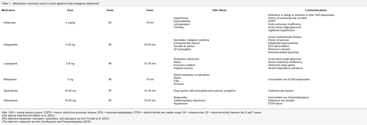
MS. M, A 64-YEAR-OLD Caucasian woman presents to the emergency department (ED) at a comprehensive stroke center with two months of intermittent loss of vision in the right eye. She complain of a headache in the right temporal area during the episodes of vision loss. Ms. M state that symptoms resolved on their own and lasted approximately 20 min. Currently, she has a headache on the right temporal region that she rates 5 out of 10 on the pain scale. Ms. M's optometrist referred her to the ED after her annual eye examination. Her eyes were still dilated from the eye examination, so vision was still blurry in both eyes. Based on her symptoms, her optometrist told her to come to the ED immediately to rule out giant cell arteritis (GCA). Her paperwork from the optometrist stated that she had a normal examination and intraocular pressures were 10 mmHg bilaterally. Associated symptoms generalized body aches, fatigue, lightheadedness, and nausea. She denied fever, chills, weight changes, jaw pain when chewing, double vision, new onset of seeing floaters or flashing lights, numbness, tingling, or painful rash to the affected side of the face. She also denied any alleviating or aggravating factors, denied pain radiating to other areas, denied previous treatment and denied any trauma or injury to the affected side.
Her medical history included prediabetes and hyperlipidemia. She denied a medical history of GCA, polymyalgia rheumatica (PMR), stroke, or migraines. Ms. M had cataract surgery in both eyes one year ago. She taking atorvastatin 20 mg PO daily. She denied taking over-the-counter medications or supplements. She had no known drug allergies. She also denied alcohol or tobacco use. Ms. M denied the use of methamphetamines in the past two years. She stated she has been sober for two years.
The patient had a body mass index of 30 kg/m2, blood pressure of 140/86 mmHg on the right arm and 145/80 mmHg on the left arm, heart rate 100 beats per minute, temperature 37.8 °C, and oxygen saturation 98% on room air.
On physical examination, the abnormality was tenderness upon palpation patient's over the right temporal region. Her eyes were still dilated bilaterally from her visit with the optometrist. Extraocular movements were intact bilaterally. Her head was normocephalic and atraumatic. Neurological examination showed cranial nerves II–VII intact, sensation intact throughout, alert, and oriented to self/place/time/situation. Temporal, radial, dorsalis pedis pulses 2+ and equal bilaterally. Capillary refill less than 3 seconds S1 and S2 sounds heard was murmurs. No carotid bruits bilaterally. The patient had clear lung sounds throughout without wheezing or crackles. Skin intact without rashes, lesions, erythema, was edema.
Complete blood cell count with differential and basic metabolic panel (BMP) was ordered and results are pending. Erythrocyte sedimentation rate (ESR) was added to blood work as well. Color Doppler ultrasonography (US) of both temporal arteries was ordered.
PATHOPHYSIOLOGY
The etiology of GCA is unknown and involves inflammation of vessels such as the aorta, carotid, temporal, or axillary arteries (Langford et al., 2017). The arteries of the scalp, head, or temples are frequently inflamed, so the term “temporal arteritis” is commonly used (Bolster, 2019). Polymyalgia rheumatica may overlap with GCA and can occur in up to 50% of patients with GCA, which makes it challenging to differentiate between the two (Bolster, 2019; Jhun, Aguilera, Shoenberger, Briht, & Herbert, 2015). PMR is an inflammatory condition associated with an elevation of ESR and C-reactive protein (CRP), but it is characterized by pain and stiffness in proximal muscles (Sharma, Mohammad, & Turesson, 2020). Giant cell arteritis does have a diagnostic criterion and the treatment is similar to PMR. The difference is GCA requires a higher dosage of corticosteroids than PMR.
In patients with GCA, the goal is to improve symptoms and prevent permanent blindness (Langford et al., 2017). Other complication that can occur is scalp or tongue necrosis or Raynaud's phenomenon in the upper extremities, due to decreased blood flow to these areas, but this is rare (Ameer, Peterfy, & Khazaeni, 2021). Cerebrovascular events can occur as well such as transient ischemic attacks or stroke (Ameer et al., 2021). Symptoms include worsening headache, visual changes (blindness or blurry), tongue/jaw pain, pain in extremities, scalp tenderness, peripheral or mononeuropathy, weakness, weight loss, or fatigue (Ameer et al., 2021; Langford et al., 2017). Physical examination might reveal fever, diminished or absent temporal artery pulse (unilateral), scalp tenderness, or temporal artery tenderness with localized erythema and edema (Langford et al., 2017). Performing an eye examination using the ophthalmoscope is important to evaluate for ischemic optic neuropathy. If acute ischemic optic neuropathy is present, then there will be a swollen pale disc in the affected eye (Hellmich et al., 2020). If the inflammation is in the aorta or carotid arteries, then bruits in carotid arteries or asymmetric arm blood pressure readings may be present (Wojczal, Kozera, Luchowski, & Neubauer-Geryk, 2019).
EPIDEMIOLOGY
After the age of 50 years, the incidence of GCA increases, reaching a peak incidence in the age group of 71–80 years (Wojczal et al., 2019). According to Sharma et al. (2020), GCA is more common in women than in men with an approximate ratio of 3:1. Around 40%–60% of patients diagnosed with GCA have been reported to have signs and symptoms of PMR (Sharma et al., 2020).
Diagnostic findings:
American College of Rheumatology (ACR)
European Alliance of Associations for Rheumatology (EULAR)
Diagnostic criteria
Three out of five criteria that need to be present include signs/symptoms and diagnostic findings (Hunder et al., 1990)
Categorized as active disease, relapse (minor and major), and remission based on presence of any signs/symptoms and one diagnostic finding (Hellmich et al., 2020)
Signs and symptoms
Disease onset at 50 years of age or older
Temporal artery tenderness to palpation or decreased pulsation
New-onset headache or new type of localized pain in the head (Hunder et al., 1990)
New onset of persistent localized headache
Constitutional symptoms (weight loss of more than 2 kg, low-grade fever, fatigue, night sweats)
Jaw and/or tongue pain when chewing
Temporary loss of vision or double vision
Symptoms of PMR
Limb pain when ambulating or using arms
Tenderness and/or thickening of the superficial temporal arteries with or without reduced pulsation
Scalp tenderness
Bruits (particularly in the axilla)
Reduced pulses/blood pressure of the upper limbs
Anterior ischemic optic neuropathy
Oculomotor cranial nerve palsy/palsies
Central retinal artery occlusion, branch retinal artery occlusion, and/or choroidal ischemia (Hellmich et al., 2020)
Diagnostic findings
Elevated ESR (ESR of 50 mm/hr or greater by the Westergren method)
Abnormal artery biopsy showing vasculitis characterized by a predominance of mononuclear cell infiltration or granulomatous inflammation, usually with multinucleated giant cells (Hunder et al., 1990)
Current GCA activity on imaging or biopsy
Persistent elevated inflammatory markers (ESR or CRP; Hellmich et al., 2020)
Ultrasonography (US) is ideal for first imaging modality due to low-cost, noninvasive, and no radiation exposure. The results for US in a patient with GCA might show hypoechoic, noncompressible, edematous wall of the artery also known as the “halo sign” for GCA (Wojczal et al., 2019). The “halo sign” does disappear with glucocorticoid treatment after 2–3 weeks (Wojczal et al., 2019). If US is not available, then high-resolution magnetic resonance imaging (MRI) of cranial arteries can be ordered (sensitivity 69% and specificity 91%; Dejaco et al., 2018). The reason a computed tomography is not recommended to rule out GCA is due to radiation exposure and lack of evidence (Dejaco et al., 2018).
Temporal artery biopsy can be performed in the hospital or outpatient setting. However, up to 44% of patients with clinical features of GCA have a negative biopsy (Wojczal et al., 2019). It is important for providers to know that imaging or biopsy should not delay the initiation of treatment, so if GCA is highly suspected, then treatment should be started immediately (Dejaco et al., 2018). Therefore, the diagnosis of GCA can be made without biopsy or further imaging.
EVIDENCE-BASED TREATMENT GUIDELINES
If GCA is strongly suspected, then the ACR and the EULAR recommend starting treatment right away (Hellmich et al., 2020; Hunder et al., 1990). Another reason for starting treatment immediately for GCA before obtaining diagnostic tests is to prevent irreversible visual loss (Hellmich et al., 2020). When there is GCA-related visual symptoms, then the provider should consider the use of intravenous methylprednisolone 250–1,000 mg/day for 3 days and then proceed to the recommended oral prednisone schedule (Hellmich et al., 2020).
Once the disease is controlled, the recommended prednisone schedule is tapering to a target dose of 15–20 mg/day within 2–3 months, and after 1 year to 5 mg/day or less (Hellmich et al., 2020). The ACR does recommend different dosing for the oral prednisone schedule. According to the ACR, initial treatment consists of prednisone 40–60 mg/day for at least a month and then it can be tapered slowly (Bolster, 2019).
Usually, symptoms resolve quickly with treatment, but during the tapering process, GCA symptoms can recur (Bolster, 2019). The tapering dose can be reduced to 5–10 mg/day over a few months (Bolster, 2019). However, tapering process can take 1–2 years to prevent recurring GCA (Bolster, 2019). Most patients are able to discontinue corticosteroid use after a few years of the disease onset, but some may require long-term use of low-dose corticosteroids (Ameer et al., 2021). GCA does not affect the overall survival rate of an individual except for those with aortic involvement (may lead to abdominal aortic aneurysm or dissection; Ameer et al., 2021). Adverse effects of treatment include bone loss from chronic corticosteroid usage, which could lead to osteoporosis or fractures (Ameer et al., 2021). Therefore, additional management might be necessary such as bone density scanning and treatment with bisphosphonates (Ameer et al., 2021).
Recently, tocilizumab was approved for the treatment of GCA in conjunction with prednisone and allowed for less prednisone to be prescribed (Bolster, 2019). According to the EULAR, adjunctive therapy should be used in patients with refractory or relapsing GCA using tocilizumab or methotrexate (Hellmich et al., 2020). Tocilizumab can be given monthly intravenously or subcutaneously every week or two (Bolster, 2019). Depending on the dose, this medication can cost anywhere from $100 to $20,000, so this may not be easily accessible to all patients (Bolster, 2019). Finally, the EULAR does not recommend antiplatelet or anticoagulant therapy for patients with GCA unless it is indicated for other reasons (Hellmich et al., 2020).
ED MANAGEMENT
When a patient presents to the ED with suspected GCA, it is crucial that a thorough history and physical examination is performed to determine whether glucocorticoid therapy needs to be started prior to diagnostic testing. If the patient with suspected GCA is having visual disturbances, the ENP will need to start intravenous methylprednisolone 250–1,000 mg/day prior to diagnostic testing to prevent permanent blindness (Hellmich et al., 2020). In addition, appropriate consults need to be called and patient will need to be admitted inpatient to continue intravenous therapy (Hellmich et al., 2020). Consults should be made to rheumatology, for the inflammatory disorder; ophthalmology, for visual disturbances; and neurology, for neurological findings. Once treatment has been started and consults called, then diagnostic testing can be completed (Hellmich et al., 2020). In the ED or outpatient setting, color Doppler US of the temporal arteries should be the first imaging modality (sensitivity 67% and specificity 91%; Dejaco et al., 2018). Appropriate diagnostic testing includes US of the temporal or axillary arteries to look for the “halo sign” on the affected side. If US is not easily accessible, then MRI can be ordered. Labs such as ESR or CRP should be ordered as well. Temporal artery biopsy is completed inpatient or outpatient, so this would not be completed in the ED.
When a patient with suspected GCA without visual disturbances presents to the ED, then management can be completed outpatient. Diagnostic testing such as imaging and blood work can be completed in the ED to help support the diagnosis of GCA or it can be completed outpatient. It is crucial that the ENP informs the patient the importance of following up outpatient for the management of GCA. Oral glucocorticoid therapy should not be started in the ED; instead, it should be managed outpatient by a specialist.
The ENP needs to educate the patient with GCA and ensure patient understanding on when to return to the ED. Patients with GCA should return to the ED immediately for fever, worsening headache, change in speech, change in sensation on face or extremities, or visual disturbances (blurry vision, double vision, blindness).
OUTPATIENT MANAGEMENT
Outpatient management consists of long-term glucocorticoid therapy and monitoring of GCA relapse by a rheumatologist (Wojczal et al., 2019). If a biopsy, imaging, or blood work has not been conducted in the hospital, then it can be performed outpatient. Patients need to be educated on the importance of attending follow-up appointments to assess medication management and possible relapse of GCA.
DIFFERENTIAL DIAGNOSIS
Diagnosing GCA is mostly based on the patient history and physical examination. Given the patient's history of headache, unilateral vision loss, and tenderness of right temporal artery without focal neurological deficits, presentation is concerning for temporal arteritis. Complex migraine is still a possibility, but the patient does not have a history of migraines. The patient denies recent trauma or injury, so there is low suspicion for corneal abrasion, corneal ulcer, or orbital fracture. There is low suspicion for acute angle-closure glaucoma due to normal intraocular pressure. Herpes zoster ophthalmicus is unlikely because the patient does not have a painful rash on the affected side. There is low concern for cerebrovascular accident or transient ischemic attack because the patient does not have focal neuro deficits. Low concern for retinal detachment because the patient denied seeing floaters or flashing lights.
REVISITING THE CASE
Based on Ms. M's signs and symptoms (visual disturbances, headache, and temporal artery tenderness), the ED provider highly suspected GCA. Also, Ms. M's age and sex place her at a high risk for GCA. To confirm active GCA, the ED provider ordered blood work and imaging. Ms. M's laboratory test results revealed an elevated ESR at 100 mm/hr. Complete blood cell count with differential and BMP was within normal limits. US of both temporal arteries showed hypoechoic circumferential thickening around the right temporal artery lumen; left temporal artery was unremarkable.
According to the ACR criteria, Ms. M meets at least three criteria for active GCA. Ms. M is 50 years of age of older (64 years), has temporal artery abnormality (right temporal artery tenderness to palpation), and has ESR of 50 mm/hr or greater (100 mm/hr).
Ms. M meets several of the EULAR criteria for active GCA such as disease onset at 50 years of age or older (64 years), constitutional symptoms (lightheadedness and nausea), acute visual symptoms (intermittent unilateral loss of vision), symptoms of PMR (generalized body aches and fatigue), ESR of 50 mm/hr or greater (100 mm/hr), and current GCA activity on imaging (US result “halo sign” right temporal artery; Hellmich et al., 2020).
Currently, Ms. M does not have vision loss, but it is difficult to perform an accurate ophthalmology examination due to her eyes being dilated from her annual eye examination. The ED provider decided to consult rheumatology first to determine whether or not intravenous methylprednisolone should be started in the ED. The major concern was recurring unilateral vision loss potentially leading to permanent blindness. Based on the patient's reported history of intermittent vision loss, the rheumatologist advised the ED provider to start methylprednisolone 1 g intravenously daily and admit the patient to inpatient telemetry. Additional consults were placed for neurology and ophthalmology.
CONCLUSION
Giant cell arteritis can mimic signs and symptoms of other disease processes, so it is important for the ENP to be knowledgeable in the diagnostic criteria presented by the ACR or the EULAR. It is crucial for ENPs to recognize and start immediate treatment in patients with suspected GCA plus visual disturbances to prevent further complications such as permanent blindness. Finally, ENPs need to emphasize the importance of following up outpatient for patients with GCA to avoid further complications or relapse.
REFERENCES
Ameer M. A., Peterfy R. J., Khazaeni B. (2021). Temporal arteritis. Treasure Island, FL: StatePearls Publishing. Retrieved November 14, 2020, from
https://www.ncbi.nlm.nih.gov/books/NBK459376/
Bolster M. (2019). Giant cell arteritis. Atlanta, GA: American College of Rheumatology. Retrieved November 13, 2020, from
https://www.rheumatology.org/I-Am-A/Patient-Caregiver/Diseases-Conditions/Giant-Cell-Arteritis
Dejaco C., Ramiro S., Duftner C., Besson F. L., Bley T. A., Blockmans D., Schmidt W. A. (2018). EULAR recommendations for the use of imaging in large vessel vasculitis in clinical practice. Annals of the Rheumatic Diseases; London, 77(5), 636. doi:10.1136/annrheumdis-2017-212649
Hellmich B., Agueda A., Monti S., Buttgereit F., de Boysson H., Brouwer E., Luqmani R. A. (2020). 2018 Update of the EULAR recommendations for the management of large vessel vasculitis. Annals of the Rheumatic Diseases, 79(1), 19–30. doi:10.1136/annrheumdis-2019-215672
Hunder G. G., Bloch D. A., Michel B. A., Stevens M. B., Arend W. P., Calabrese L. H., Lie J. T. (1990). The American College of Rheumatology 1990 criteria for the classification of giant cell arteritis. Arthritis and Rheumatism, 33(8), 1122–1128. doi:10.1002/art.1780330810
Jhun P., Aguilera P., Shoenberger J., Briht A., Herbert M. (2015). Giant cell arteritis: Read the fine print! Annals of Emergency Medicine, 65(5), 615–617. doi:10.1016/j.annemergmed.2015.02.020
Langford C. A., Cuthbertson D., Ytterberg S. R., Khalidi N., Monach P. A., Carette S., Merkel P. A. (2017). A randomized, double-blind trial of Abatacept (CTLA-4Ig) for the treatment of giant cell arteritis. Arthritis & Rheumatology, 69(4), 837–845. doi:10.1002/art.40044
Sharma A., Mohammad A. J., Turesson C. (2020). Incidence and prevalence of giant cell arteritis and polymyalgia rheumatica: A systematic literature review. Seminars in Arthritis and Rheumatism, 50(5), 1040–1048. doi:10.1016/j.semarthrit.2020.07.005
Wojczal J., Kozera G., Luchowski P., Neubauer-Geryk J. (2019). Advantages in diagnosis of giant cell arteritis by ultrasound. Advances in Dermatology and Allergology, 36(1), 25–28. doi:10.5114/ada.2019.82823


Comments (0)