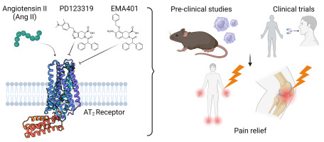
NMR applications to GPCR recognition by peptide ligands
G-protein coupled receptors (GPCR) are the largest superfamily of membrane proteins with more than 800 members that transduce signals from a wide range of stimuli including small molecules, lipids, peptides, and proteins [1]. Among which peptides are the most prevalent endogenous ligands activating GPCRs across the entire family. To date more than 180 principal peptide ligands have been paired with more than 120 GPCRs, and are increasing in number as deorphanization progresses [2]. Although GPCRs are targeted by more than 30% of FDA approved drugs, only a small percentage of marketed drugs act via peptide GPCRs [3]. This limitation is likely due to the general technical problems in purifying GPCRs and the complicated mode of binding of peptides to GPCRs that hampers rational and structure-based drug design approaches.
In the past decade technological advances in the expression and purification of GPCRs along with improvements in crystallography and cryo-EM methods have enabled solution of the structure of more than 40 unique peptide GPCRs, mainly in complex with non-endogenous ligands [4]. Although these snapshots have revealed the main aspects of receptor conformational changes upon activation, the mechanism by which peptides recognize the receptor and shift the conformational equilibrium has remained elusive. In contrast to small molecule ligands, such as adrenaline and adenosine, peptides are highly dynamic even in the bound state, tend to bind to shallower pockets, make extensive networks of interactions with the extracellular loops (ECLs) of the cognate receptor and usually undergo conformational changes upon binding. These conformational changes range from slight reorganization of backbone and side chains to remarkable large amplitude secondary structure transitions that can modify the dynamic landscape of the peptide ligand over the whole range of timescales from the very slow secondary structure rearrangement to the very fast side chain rotations. Indeed, the slow timescale motion of peptides is likely to fine tune the receptor response to the ligand [5] and the very fast entropically favorable side-chain motions may contribute to the relatively high affinity of peptide ligands compared to small molecule ligands [6]. Whether the peptides undergo conformational selection, in which the folding occurs prior to binding, or induced fit, in which folding is preceded by the binding event, the mechanism of recognition and binding of peptides to GPCRs remains an important question. While the static cryo-EM and X-ray crystallography techniques are limited in investigating biomolecular dynamics, spectroscopy techniques, including NMR, are well suited to investigate such processes at atomic resolution. While NMR is the method of choice to study formation of encounter complexes and the conformational changes that are essential for steering the ligand to the orthosteric binding pocket, in combination with other techniques, such as stopped-flow fluorescence [7,8], surface plasmon resonance [9], biolayer interferometry [10] and molecular dynamic simulations [11], increases the depth of understanding of the kinetic pathways of receptor recognition. In this review, we will highlight the importance of binding induced folding of intrinsically disordered peptide ligands in receptor recognition and the applicability of NMR techniques to tackle this question.


Comments (0)