
Remember me
Cardiovascular diseases (CVD) are still a leading cause of morbidity and mortality worldwide,1 among which acute ST elevation myocardial infarction (STEMI) contributes to a substantial number of these events, with immediate reperfusion by either primary percutaneous intervention (p PCI) or thrombolytic agents being the most effective treatment strategy adopted nowadays.2
Over the last 30 years, nuclear cardiology has become a mainstay in the evaluation of ischemic heart diseases both on the diagnostic and prognostic aspects in the setting of acute coronary syndromes (including myocardial infarction or unstable angina).3
Osteopontin (OPN), a member of the small integrin-binding ligand n-linked glycoprotein (SIBLING) family that was initially discovered as a bone matrix protein, is currently implicated in a variety of acute as well as chronic inflammatory processes, including atherosclerosis and it has been recently found to be an important player in ischemic vascular pathologies including myocardial infarction.4
The fact that acute STEMI patients even with similar demographics, risk factors and treatment modality end up with diverse outcomes as regards cardiac functionality, emphasizes the need for prognostic tools to help identify subjects at higher risk of suffering adverse outcome post STEMI which might help to improve targeted treatment and follow-up strategies for those patients.
2 Patients and methodsTo investigate the role of serum OPN in prediction of short-term outcome (within one month) in STEMI patients, all patients presenting with their first STEMI to the critical care department at Cairo University hospitals, during the period from January 2014 till December 2014 were prospectively recruited for the study and compared to matched control subjects with normal coronary angiography. All patients received an informed written consent, and the study protocol was approved by the local review board and ethical committee. Evidence of new STEMI required presence of chest pain for more than 30 minute and <12 hours and cumulative persistent new ST elevation at the J point in at least 2 contiguous leads of ≥2 mm (0.2 mV) in men or ≥1.5 mm (0.15 mV) in women in leads V2–V3 and/or of ≥1 mm (0.1 mV) in other contiguous chest leads or the limb leads. New or presumably new left bundle branch block has been considered a STEMI equivalent.5 Exclusion criteria included patients with STEMI of more than 12 hours from presentation, Patients with UA/non-STEMI, patients previously known to have a significant multivessel disease, patients having any co-morbid disease that affects life expectancy as terminal malignancies, also chronic liver or kidney disease and connective tissue disorders associated with elevated OPN level were excluded as well.
All the studied population were subjected to full history taking, detailed clinical examination, 12 lead ECG, Cardiac biomarkers (CK, CKMB, troponin T) assessment and Serum OPN level was assessed on admission, All patients with STEMI underwent revascularization with primary PCI, myocardial perfusion imaging to assess the infarction size (IS) after 3 days of admission, an echocardiography on admission and follow-up after 1 month together with monitoring occurrence of MACE (left ventricular [LV] systolic dysfunction heralded by ejection fraction (EF) < 40%,6 reinfarction, malignant arrhythmias, stroke, or death).
Data were statistically described in terms of mean ± standard deviation (±SD), median and range, or frequencies (number of cases) and percentages when appropriate. Comparison of numerical variables between the study groups was done using Student t test for independent samples in comparing 2 groups when normally distributed. Correlation between various variables was done using Pearson moment correlation equation for linear relation in normally distributed variables and Spearman rank correlation equation for non-normal variables/non-linear monotonic. Receiver operating characteristic curve was used to detect the cutoff value. P < .05 was considered statistically significant. All statistical calculations were done using computer program SPSS (Statistical Package for the Social Science; SPSS Inc., Chicago, IL) release 15 for Microsoft Windows (2006).
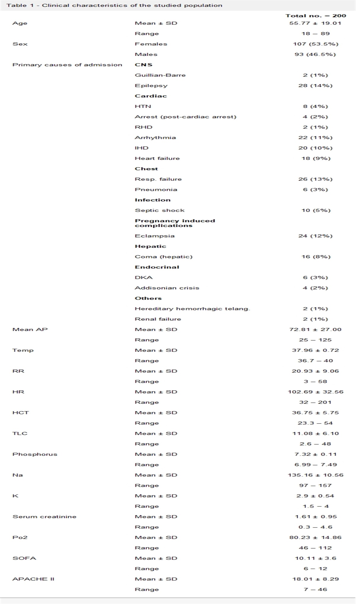
3 ResultsForty consecutive patients with STEMI (29 males [73%], mean age [51.3 ± 11.2 years]) were enrolled in the study, designated as Group 1 (Cases) (Tables 1 and 2). They were compared to 30 matched controls with normal coronary angiography (19 males [63%], mean age [48.4 ± 9.2 years]) Group 2 (Control).
Table 1 - Risk factors for STEMI Risk factors Number of patients (%) Diabetes mellitus 16 40 Hypertension 18 45 Smoking 30 75 Dyslipidemia 22 55 Family history 15 37.5STEMI, ST elevation myocardial infarction.
LV, left ventricular; MACCE, major adverse cardiac and cerebrovascular events.
Forty percent of the studied patients were diabetics, 45% were hypertensives, 75% were smokers, 55% were dyslipedemic while 37.5% were found to have family history of coronary artery disease.
3.1.2 Time of presentationThe time of presentation from the onset of chest pain varied from 5 to 12 hours with a mean of 8.25 hours (8.25 ± 2.7 hours). All patients underwent primary PCI with mean door to balloon time (60 ± 30 minutes).
3.1.3 Electrocardiographic dataAccording to the electrocardiographic (ECG) findings in group 1 (patients), the site of STEMI was anterior in 22 patients and inferior in 18 patients.
3.1.4 OPN levelIn the current study, group 1 patients showed mean OPN level (106.9 ± 15 ng/dL), while in group 2 (control), the mean level (27.5 ± 12.7 ng/dL) with group 1 showing a higher level with strong statistical difference (P .01) (Fig. 1).
 Figure 1:
 Serum OPN level in cases and control groups.
3.1.5 Echocardiographic data (ejection fraction)In our study, group 1 patients showed an EF ranged from 33% to 67% with Mean EF (49.67% ± 9.9%).
3.1.6 Nuclear imaging data (infarction size)In our study, the IS ranged from 6% to 42% with mean 21.8% in group (1) patients
3.1.7 Complications of STEMI (major adverse cardiac and cerebrovascular events)Twenty-four STEMI patients (60%) developed major adverse cardiac outcomes.
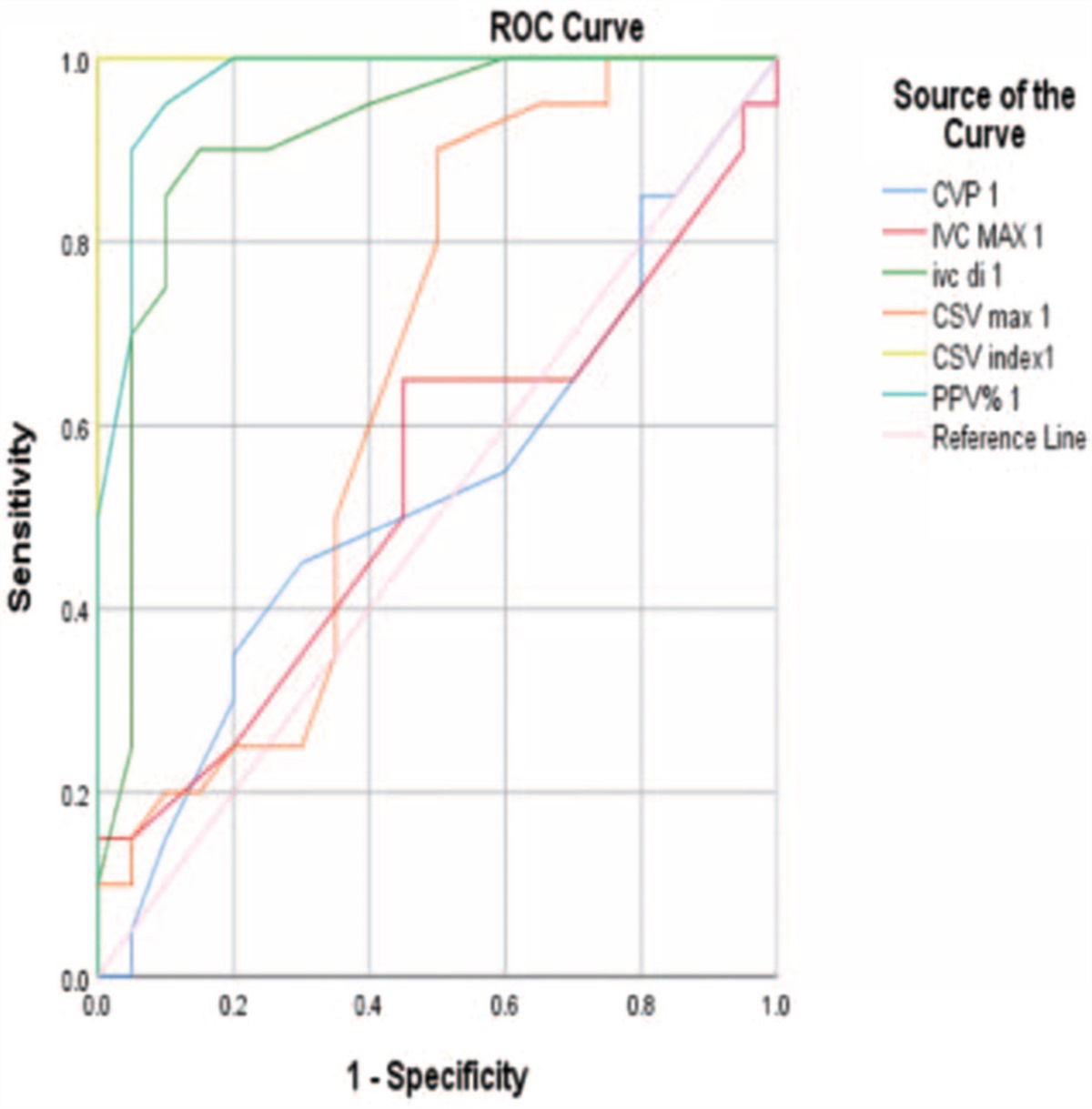
3.2 Correlations 3.2.1 OPN and LV systolic dysfunctionThere was a statistically significant correlation between serum OPN level and LV systolic dysfunction in group 1 within one month of follow-up (r: 0.3, P: .045). Our study suggests a cutoff value of (104 ng/dL) as a potential predictor for LV systolic dysfunction (Figs. 2 and 3).
 Figure 2:
 Showing the relation between serum OPN level and the LVEF. LVEF, left ventricular ejection fraction.
 Figure 3:
 ROC curve, cutoff value of OPN for prediction of LV systolic dysfunction. LV, left ventricular; ROC, receiver operating characteristic.
While on the other hand, there was no significant correlation between OPN level and mortality (r: 0.1, P: .8), reinfarction (r: 0.1, P: .9) or occurrence of malignant arrhythmias (r: 0.2, P: .8).
3.2.2 IS and correlations with other variablesThere was a statistically significant correlation between IS and LV systolic dysfunction in group 1 during short term follow-up (r: 0.9, P: .0001). Figure 4 demonstrates the negative linear relation between IS and EF.
 Figure 4:
 The relation between IS and EF. EF, ejection fraction; IS, infarction size.
3.3 IS and mortalityThere was a statistically significant correlation between IS and mortality in group 1 during short term follow-up (r: 0.6, P: .003).
However, there was no correlation between IS and malignant arrhythmias (r: 0.2, P: .5) or reinfarction (r: 0.7, P: .85).
3.4 IS and OPNThere was a statistically significant positive correlation between serum OPN level and IS (r: 0.31, P: .045) (Fig. 5).
 Figure 5:
 Relation between serum OPN level and infarction size.
4 DiscussionTo the best of our knowledge, the present study addresses, for the first time the impact of serum OPN on short-term clinical outcome in STEMI patients treated with pPCI.
Serum OPN level was significantly higher in STEMI patient than in normal individuals. In addition, high OPN level independently predicted LV systolic dysfunction, but it had no significant correlation with reinfarction, malignant arrhythmias, stroke, or death.
Our data was similar to those published by Tamura et al.7 who detected an increase in serum OPN level following myocardial infarction, and a positive correlation between high OPN level and left-ventricle remodeling; The authors suggested that OPN could be used as a serum marker of ventricular remodeling and hence LV systolic dysfunction.
Similarly, in another study, conducted by Suezawa et al.,8 they demonstrated time-dependent changes in the plasma level of OPN and found that these changes are correlated with LV volumes and function, indicating that the plasma level of OPN may provide clinically helpful information for the evaluation of the pathologic processes of infarction, thus might be used as an indicator of LV remodeling after AMI. Also that study revealed a positive correlation between the plasma OPN level and the LV end-systolic volume index, and a negative correlation with the left ventricular ejection fraction (LVEF).
In concordance with our study, M. Singh et al.9 studied the role of OPN in extracellular matrix protein deposition and myocardial remodeling post MI, and they found that OPN expression increases in the heart after MI, and this has the potential to increase cardiac fibrosis leading to LV dysfunction.
Our results revealed that there was a statistically significant correlation between IS determined by myocardial perfusion imaging and LV systolic dysfunction in group 1 during 1 month follow-up period, and that there was a significant negative linear relationship between infarct size and LVEF especially for those with larger IS (P: .0001, r: 0.9).
As regard the correlation between IS and mortality, there was a significant correlation especially for those who had larger IS (P: .003, r: 0.6).
However, there was no correlation between IS and malignant arrhythmias (r: 0.4, P: .94) or reinfarction (r: 0.5, P: .85).
In concordance with our results, B. Pride et al.10 studied the relation between IS in STEMI patients treated successfully by p PCI and LVEF at 3 months after the infarction, they concluded that there was a negative linear relation between infarct size and LVEF for moderate to large infarcts. For small infarctions there was no significant relation between infarct size and LVEF.
Our data had also been similar to another study conducted by Burns et al.11 who have demonstrated an association between IS after successfully treated STEMI and 6-month mortality in a much larger series of 1184 patients.
Finally, in the detailed analysis of the primary findings of the present study compared with the published literature, STEMI patients had different outcome despite similar risk factors, presentation and management, so different tools should be used to predict their outcome, based upon our study, we do recommend using of serum OPN level on admission of patients with acute STEMI to aid in achieving this goal by detecting patients at risk to develop LV dysfunction in order to intensify follow-up and enhance their awareness of the warning signs of heart failure.
Also, IS determination by MPI could similarly be of value in predicting LV dysfunction and mortality in STEMI patients.
4.1 The study limitationsSmall number of patients due to financial constraints, may decrease the power of the study. Further larger studies are warranted to confirm the validity of our interesting findings.
5 ConclusionSerum OPN level might be a useful prognostic short-term marker in patients with STEMI after revascularization (regarding LV systolic dysfunction only). Similarly, OPN correlated with IS, where the latter is a good predictor for LV systolic dysfunction and mortality.
References [1]. Lloyd-Jones D, Adams R, Carnethon M, et al. Heart disease and stroke statistics-2009 update: a report from the American Heart Association Statistics Committee and Stroke Statistics Subcommittee. Circulation 2009;119:e21–e181. [2]. Roe MT, Messenger JC, Weintraub WS, et al. Treatments, trends, and outcomes of acute myocardial infarction and percutaneous coronary intervention. J Am Coll Cardiol 2010;56:254. [3]. Kim SC, Adams SL, Hendel RC, et al. Role of nuclear cardiology in the evaluation of acute coronary syndromes. Ann Emerg Med August 2007;30:210–218. [4]. Ohmori R, Momiyama Y, Taniguchi H, et al. Plasma osteopontin levels are associated with the presence and extent of coronary artery disease. Atherosclerosis 2003;170(2):333–337. [5]. Thygesen K, Alpert JS, Jaffe AS, et al. Third universal definition of myocardial infarction. Eur Heart J 2012;33:2551–2567. [6]. Lindenfeld J, Albert NM, et al. Heart Failure Society of America. HFSA 2010 comprehensive heart failure practice guideline. J Card Fail 2010;16:e1–e194. [7]. Tamura A, Shingai M, Aso N, et al. Osteopontin is released from the heart into the coronary circulation in patients with a previous anterior wall myocardial infarction. Circ J 2003;67:742–744. [8]. Suezawa C, Kusachi S, Murakami T, et al. Timedependent changes in plasma osteopontin levels in patients with anterior-wall acute myocardial infarction after successful reperfusion: correlation with left ventricular volume and function. J Lab Clin Med 2005;145:33–40. [9]. Singh M, Foster CR, Dalal S, Singh K. Role of OPN in extracellular matrix deposition and myocardial remodeling post-M. J Mol Cell Cardiol 2010;48:538–554. [10]. Pride YB, Giuseffi JL, Mohanavelu S, et al. Relation between infarct size in ST-segment elevation myocardial infarction treated successfully by percutaneous coronary intervention and left ventricular ejection fraction three months after the infarct. Am J Cardiol 2010;106:635–640. [11]. Burns RJ. LV function and infarct size predict six-month mortality post MI treated by thrombolysis. Circulation 2004;94(suppl I):I–655.
Comments (0)