Plant collection and authentication
Sclerocarya birrea fruit samples were collected at coordinates ‘22°35′35.7″ S 27° 07′ 28.5″ E’, which is in the township of Palapye, located in the Central district of Botswana. The plant was authenticated at the Herbarium laboratory at University of Botswana, voucher number BAK-02/2020.
Sample preparation
Sclerocarya birrea fruits were pilled to separate the exocarp from the mesocarp after being rinsed with distilled water. The fruit exocarp was cleaned with distilled water before being dried for 48 h in a Lasec MMMERT UF 110 laboratory oven at 40 °C. Using a Salton SB400E blender, the dried materials were crushed into a powder before being sequentially extracted in Hexane, Chloroform, Ethyl acetate, and Methanol as prescribed by Mustafa et al., (2020) with minor changes. In round bottom flasks, precisely 300 g of dry powder were soaked in 1 L of hexane for 76 h before being filtered through Whatman filter paper, Grade 1 (Merck, South Africa). Following a 6-h air drying period, the plant residue was once more immersed in 1 L of chloroform for 76 h. The procedure was repeated using ethyl acetate and methanol.
Determination of total flavonoid content (TFC)
The aluminum chloride colorimetric assay was used to calculate the TFC of the fruit exocarp of Sclerocarya birrea as described by Sembiring et al. (2018). In separate tubes, an aliquot (0.5 mL) of the extract and standard solution of quercetin (0.0312, 0.0625, 0.125, 0.25, 0.5,1 mg/mL) was obtained. 2 mL of distilled water and 0.15 mL of sodium nitrite (5% NaNO2, w/v) were then added, and the mixture was allowed to stand for 6 min. After incubating for 6 min, 0.15 mL aluminum chloride (10% AlCl3) and 2 mL sodium hydroxide (4% w/v, NaOH) were added, and the volume was then increased to 5 mL with distilled water. The liquid turns pink after 30 min of incubation, and a Thermo Scientific TM SPECTRONICTM 200 Spectrophotometer was used to determine its absorbance at 420 nm. Distilled water was used as a blank. Every analysis was done in triplicates, represented as mg quercetin equivalent (QE)/g material, and the TFC was calculated as
where c = concentration of extract equivalence to that of quercetin (quercetin standard curve), V = volume of extracts in mL and m = mass of extracts in grams.
Determination of total phenolic content (TPC)
Sclerocarya birrea's TPC was calculated utilizing a technique used by (Singleton et al. 1999). 200 µL of 10% (v/v) Folin-Ciocalteu reagent, 2 mL of distilled water, and an aliquot (0.2 mL) of the extract or standard solution of gallic acid (0.0312, 0.0625, 0.125, 0.25, 0.5, 1 mg/mL) were mixed and incubated for 8 min in the dark. 600 µL of 20% (w/v) sodium carbonate was then added, and the mixture was incubated for 2 h at room temperature in the dark. Every analysis was done in triplicates. Thermofisher Scientific, USA's Spec200E UV-V spectrophotometer was used to measure the amount of absorbance at 760 nm. The data for gallic acid was used to plot a standard curve (y = 0.12 + 0.37x, R2 = 0.99) that was used in the analysis.
where c = concentration of extract equivalence to that of gallic acid (from gallic acid standard curve), V = volume of extracts in mL and m = mass of extracts in grams.
Antioxidants assaysDPPH assay
The method employed to ascertain the radical scavenging activity of extracts on 2, 2-diphenyl-1-picrylhydrazyl (DPPH) was modified, as described by (Lv et al. 2018). Gallic acid (1 mg/mL) and extracts (1 mg/mL) were serially diluted to achieve concentrations of 0.0312, 0.0625, 0.125, 0.25, 0.5, and 1 mg/mL. The wells of a 96-well plate received 150 µL of 0.1 M DPPH. 50 µL of plant extracts were used to treat DPPH for wells marked as treatment. A 50 µL solution of gallic acid was used to treat the DPPH in the wells selected as positive controls. DPPH was given a 50 µL methanol treatment for the wells designated as a negative control. At room temperature and in the dark, the reactions were incubated for 30 min. After 30 min, optical density (OD) was measured at 518 nm using a MULTISKAN FC microplate reader from Thermo Scientific (USA). The following is how the percentage inhibition (decolorization) was calculated:
$$ }\;} (\% ):\frac}\;} - }\;}} \right)}}}\;}}} \times 100 $$
FRAP assay
With a few changes, Berker et al. (2007)'s description of this Ferric Reducing Antioxidants Power (FRAP) was employed. 8.3 mL of hydrochloric acid (HCL) and 100 mL of distilled water were mixed to create 1.0 N HCL. Gallic acid and plant extracts were serially diluted into quantities of 0.0312, 0.0625, 0.125, 0.25, 0.5, and 1 mg/mL. A 96-well plate had 40 µL of each concentration of plant extracts, methanol only and gallic acid added to the wells. The 96 well plates received 100 µL of 1.0 N HCL. 20 µL of sodium dodecyl sulphate at 1% and 30 mL of potassium ferrocyanide at 1% came next. The reaction was allowed to sit at 50 °C for 20 min. Wells treated with gallic acid designated positive control while wells treated with methanol were used as negative control. Using a MULTISKAN FC microplate reader from Thermo Scientific (USA), absorbance was measured at 750 nm.
$$ }\;} (\% ):\frac}\;} - }\;}} \right)}}}\;}}} \times 100 $$
ABTS assay
The 2,2′-azino-bis (3-ethylbenzothiazoline6-sulfonic acid (ABTS) approach was applied in accordance with Kooy et al. (2016)'s instructions. This technique is based on ABTS radical cation decolorization. To make the ABTS working solution, 100 mL of 7 mM ABTS and 100 mL of 2.4 mM potassium persulfate were combined (1:1) and left at room temperature for 16 h in the dark. 1 mL of ABTS solution was gradually diluted with methanol until an absorbance of 0.706 ± 0.01 was attained at 734 nm. Then, extracts and gallic acid concentrations of 0.0312, 0.0625, 0.125, 0.25, 0.5, and 1 mg/mL were added, and methanol was added to wells used as negative control, followed by 1 mL of the ABTS solution. The reaction was mixed and then allowed to sit for 7 min at room temperature with no light. Thermo Scientific's MULTISKAN FC microplate reader (available in the United States) was used to detect absorbance at 734 nm. The standard was gallic acid (positive control). This is how percentage inhibition was calculated:
$$ }\;} (\% ):\frac}\;} - }\;}} \right)}}}\;}}} \times 100 $$
Total antioxidants capacity (TAC) using phosphomolybdenum
TAC is an assay that is based on the formation of a stable green phosphate-Mo(V) complex as an outcome of the electron reduction of Mo (VI) to Mo(V), as stated by (Sharadanand Phatak et al. 2014). 1 mL of phosphomolybdenum (made by combining 31.4 mL of 0.6 M H2SO24, 397.49 mg of 28 mM sodium phosphate, and 494.36 mg of 4 mM ammonium molybdate) was mixed with around 0.1 mL of extracts and gallic acid. In an IncoCool LABOTEC incubator (LAMWORLD TECHNOLOGIES PTY LTD), the mixture was heated to 95 °C for 90 min before being cooled to room temperature. Absorbance was measured at 950 nm using a Spec200E UV-V spectrophotometer (Thermofisher Scientific, USA). Gallic acid was used to make a standard (y = 0.12 + 0.39x, R2 = 0.97), from which TAC of extracts was interpolated.
Cell culture studiesPreparation of plant extracts concentrations
A stock solution of 10 mg/mL of plant extracts was obtained by dissolving 100 mg of the extracts in 10 mL of 10% (v/v) dimethyl-sulfoxide (DMSO). To achieve a concentration of 400 µg/mL, 10 mL of stock solution was added to 25 mL of Dulbecco's minimum essential medium (DMEM). 400 µg/mL has been adopted from Sharadanand Phatak et al. (2014). Serial dilutions of the 400 µg/mL resulted in concentrations of 12.5, 25, 50, 100, 200, and 400 µg/mL.
Cell culture
To conduct multiple studies on the cytoprotective properties of Sclerocarya birrea fruit exocarp extracts, HeLa cells were grown. Penicillin–streptomycin (10 000 U/mL: 10 000 g/mL) and 10% of fetal bovine serum (FBS) were added to high glucose DMEM, which was used to culture HeLa cells. At 37 °C and 5% CO2, cells were incubated in a Thermo Scientific Forma SERIES 11 Water Jacket CO2 Incubator. Before being seeded into flat bottom 96-well plates, cells were counted using a Countess TM 3 Automated Cell Counter. Cell concentrations were then generated according to the output of the cell counter to achieve 1 × 10⁵ /mL.
Effects of extracts on oxidative stress biomarkers
A concentration of 1 × 10⁵/mL of 80–90% confluent HeLa cells (day 6 of passage of number 5) was treated for 2 h with 100 µL of the extracts (400 µg/mL), with untreated cells serving as the experiment's control. Using trypsin, cells were detached, then centrifuged at 1000 g for 10 min. The pellet was stirred up in cold PBS before being centrifuged at 1000 g for 10 min. The pellet was frozen for roughly 3 h at − 20 °C after being suspended in cold PBS. The pellet was frozen, thawed, frozen again, and thawed again 3 times to shatter the cells. For further investigation, the cell homogenate was kept in ice after being centrifuged at 1500 g for 10 min. Utilizing ELISA kits (Elabscience, USA) in accordance with the manufacturer's instructions, the effects of extracts on the activities of superoxide dismutase (SOD), catalase (CAT), and reduced glutathione (GSH) were assessed.
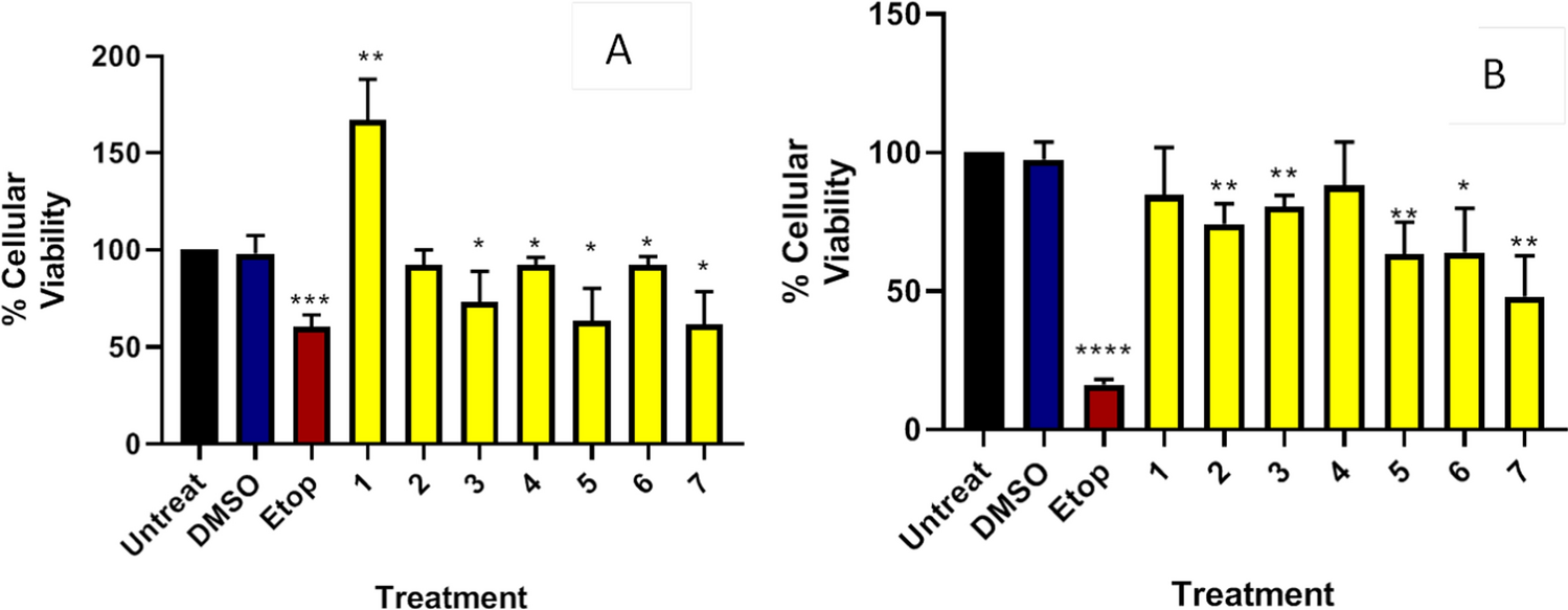
Cytoprotective effect of extracts against hydrogen peroxide-induced cell death using WST-1assay
At day 6 of passage number 5, about 90–98% of confluent HeLa cells were trypsinized, seeded in 96-well plates at a density of 1 × 10⁵/mL, and cultured for 72 h. For the wells designated as treatment, media was aspirated after incubation and replaced with 100 µL of Sclerocarya birrea fruit exocarp extracts (12.5, 25, 50, 100, 200, and 400 µg/mL). Cells in the positive control wells received PBS only. Cells were incubated for 2 h following treatment with plant extracts. By adding 10 µL of 1 mM hydrogen peroxide (H2O2) to the wells containing extracts and wells with cells treated with PBS only (positive control), and incubating for another 2 h, cells were exposed to hydrogen peroxide-induced oxidative stress. The 1 mM H2O2 was adopted from Mezghani et al. (2015). Cells were not given any treatment in negative control wells. 10 µL of (4-[-3(4-Iodophenyl)-2-(4-nitro-phenyl)-2H-5-tetrazolio]-1,3-benzene sulfonate) WST-1 was added to the wells after the incubation period and incubated for an additional 3 h. Thermo Scientific's MULTISKAN FC microplate reader (480 nm wavelength) was used to measure the absorbance. To adjust the absorbance of the treatment and control wells, background absorbance from wells containing medium and 10 µL of WST-1 was taken into account. The number of live cells that were preserved was computed as a percentage and compared to the absorbance of the control group.
$$ }\;}\;(\% ):\frac}\;}\;} - }\;}\;})}}}\;}\;})}} \times 100 $$
In silico studiesRetrieval and preparation of ligands
Phytochemicals of Sclerocarya birrea were obtained from Russo et al. (2013) and downloaded from PubChem (www.pubchem.com) as 3D structures in SDF format and were converted to PDB format using BIOVIA Discovery Studio. The Autodock tools 1.5.6 was used to add Gasteiger charges and polar hydrogens and saving each compound in pdbqt format.
Retrieval and preparation of target proteins
The 3D structure of Superoxide dismutase (PDB ID: 1CB4) and catalase (PDB ID: 2CAG) was downloaded from the Protein Data Bank (www.pdb.com). Each protein was cleaned in Autodock tools by removing water molecules, and bound ligand, and prepared by adding Kollman charges and polar hydrogens, then saved in pdbqt file.
Molecular docking
Molecular docking was performed using the Autodock 4.2 program. Briefly, the grid parameter file was prepared using Autogrid 4 in Autodock tools and grid box set at as x = 10.41, y = 87.88, z = 18.62 for SOD, and x = 58.38, y = 19.08, z = 18.3 for CAT to cover the entire catalytic sites (Rana et al. 2019). The grid box dimensions were 40 × 40 × 40 with a spacing of 0.375 angstroms. Then the docking parameter file was prepared using Autodock 4 in Autodock tools utilizing the genetic algorithm with 2,500,000 energy evaluations as the search parameter and docking using Larmakian genetic algorithms utilizing Cygwin. Docked complexes (protein + ligand) were analyzed for their interactions using BIOVIA Discovery Studio Visualizer.
Data analysis
A Graph prism software was used to analyze data and develop graphs, generated results are presented as mean ± standard error of triplicates (n = 3). To compare variation between the means at 95% confidence level (p < 0.05), we used a One way- analysis of variance (ANOVA) with Tukey–Kramer’s range test.


Comments (0)