2.1 Materials
The 20 cm × 10 cm aluminum foil-backed HPTLC silica gel 60 F254 layers (#1.05548) were acquired from Merck (Darmstadt, Germany). Analytical-grade isopropyl acetate was obtained from Sigma-Aldrich (Budapest, Hungary), and all other solvents used were of analytical grade from Molar Chemicals (Halásztelek, Hungary). Vanillin was purchased from Reanal (Budapest, Hungary). Dye reagent 3-(4,5-dimethylthiazol-2-yl)-2,5-diphenyltetrazolium bromide (MTT) was acquired from Carl Roth (Karlsruhe, Germany) and concentrated sulfuric acid (96%) from Carlo Erba (Milan, Italy). Citronella (Cymbopogon nardus) EO was obtained from a Hungarian drug store chain (Aromax Ltd., Budapest, Hungary). Test substances citronellal and citral (mixture of neral and geranial) were purchased from Sigma-Aldrich.
Gram-positive Bacillus subtilis soil bacterium (strain F1276) was a gift from József Farkas (Central Food Research Institute, Budapest, Hungary). Gram-negative, naturally luminescent marine bacterium Aliivibrio fischeri (DSM 7151) was obtained from Leibniz Institute DSMZ, German Collection of Microorganisms and Cell Cultures, Berlin, Germany, the Hungarian paprika pathogen Xanthomonas euvesicatoria from János Szarka (Primordium Kft., Budapest, Hungary) and Arabidopsis pathogen Pseudomonas syringae pv. maculicola from Jun Fan (John Innes Center, Department of Disease and Stress Biology, Norwich, UK [11]).
2.2 High-performance thin-layer chromatography
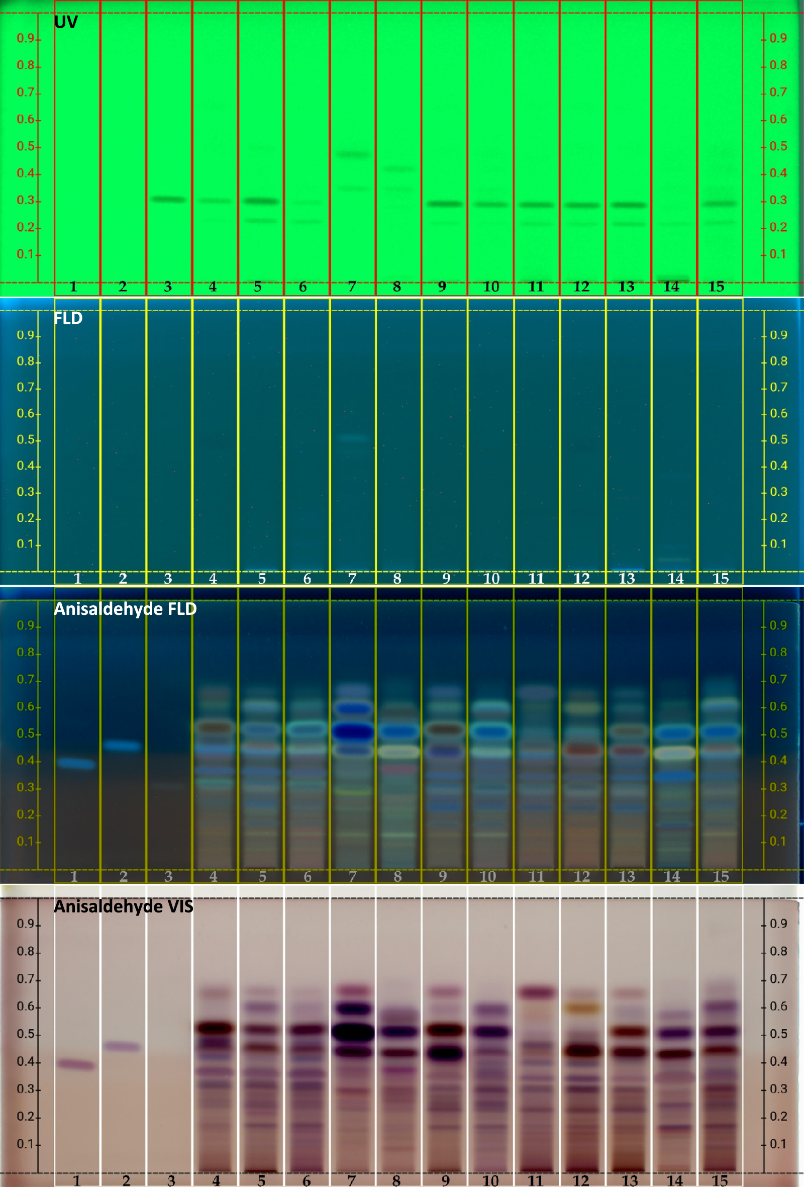
HPTLC separation was achieved in a 20 cm × 10 cm unsaturated chamber (CAMAG, Muttenz, Switzerland) with n-hexane–isopropyl acetate (9:1, v/v) as the mobile phase. CEO (30 mg/mL), citronellal (5 mg/mL), and citral (5 mg/mL) dissolved in ethanol were applied manually in the range of 2–5 µL at 8 mm height from the bottom edge in 6 mm bands by a 10 µL syringe (Hamilton, Bonaduz, Switzerland). The chromatoplates developed up to 8 cm from the lower edge were dried by a cold air stream using a hair dryer (5 min) and documented with a digital camera (Cybershot DSC-HX60, Sony, Neu-Isenberg, Germany) under an ultraviolet (UV) lamp (λ = 254 nm) (CAMAG) and at Vis after derivatization with vanillin–sulfuric acid reagent (200 mg vanillin + 50 mL ethanol + 1 mL concentrated sulfuric acid; the dipped plates were heated to 110 °C for 5 min).
For isolation, 150 μL of citronella EO solution (30 mg/mL) was applied manually as a 170 mm band by a 100 µL syringe and developed with the mobile phase n-hexane–isopropyl acetate (4:1, v/v). Then, zones of interest, determined by vanillin-sulfuric acid reagent using the left side of the chromatogram (0.5 cm), were scraped off from the remaining underivatized part into a syringe with a Teflon filter (0.22 μm, Phenomenex) and eluted with 500 μL of ethanol. The eluates were analyzed by SPME–GC–MS.
2.3 HPTLC–bioassay
The bioassays were performed using B. subtilis, A. fischeri, P. syringae pv. maculicola, and X. euvesicatoria bacterial strains based on previously published methods [8]. Briefly, the dried HPTLC plates developed for B. subtilis and X. euvesicatoria bioassays were immersed into the appropriate cell suspension, incubated for 2 h in a vapor chamber at 37 °C and 28 °C, respectively, stained with aqueous MTT solution (100 mg in 100 mL of water) by immersion, and after a 15–20 min incubation, the bioautograms were documented with the Cybershot DSC-HX60 digital camera. The bright spots against the bluish background indicate the zones of antibacterials.
In the cases of luminescent A. fischeri and P. syringae pv. maculicola, the developed layers were dipped into the cell suspensions and immediately put into a transparent glass cage under a low-light camera (iBright FL1500 Imaging System, Thermo Fisher Scientific, Budapest, Hungary). The exposure time was 40–80 s for A. fischeri and 2–3 min for P. syringae pv. maculicola. The dark zones lacking luminescent viable cells indicate antibacterial activity.
2.4 SPME–GC–MS conditions
The analysis of the EO and its compounds was carried out with an Agilent 6890N/5973N GC-MSD (Santa Clara, CA, USA) system equipped with a Supelco (Sigma-Aldrich) SLB-5MS capillary column (30 M × 250 µm × 0.25 µm). The GC oven temperature increased from 60 °C (3 min isothermal) to 250 °C at 8 °C/min (1 min isothermal). High-purity helium (6.0) was used as a carrier gas at 1.0 mL/min (37 cm/s) in constant flow mode. Static headspace solid-phase microextraction (sHS-SPME) technique was performed with an automatic multipurpose sampler (CTC Combi PAL, CTC Analytics AG, Zwingen, Switzerland) using a 65 μM StableFlex polydimethyl siloxane/carboxene/divinyl benzene (CAR/PDMS/DVB) SPME fiber (Supelco, Bellefonte, PA, USA) and 20 mL headspace vials. Extraction was performed after a 5 min incubation at 100 °C by exposing the fiber to the headspace for 10 min. Then, the fiber was immediately transferred to the injector port and desorbed for 1 min at 250 °C. Cleaning and conditioning of the SPME fiber was carried out in a Fiber Bakeout Station (Agilent) in a pure nitrogen atmosphere at 250 °C for 15 min. The mass selective detector was equipped with a quadrupole mass analyzer and was operated in electron ionization mode at 70 eV in full scan mode (41–500 a.m.u. at 3.2 scan/s). MSD ChemStation D.02.00.275 software (Agilent) was used for data analysis. Compound identification was carried out by comparing retention data and the recorded spectra with the data of the NIST 2.0 library. Percentage evaluation included area normalization.


Comments (0)