
Remember me
Thermal displays are devices that can present a desirable temperature cue to the skin. Thermal displays have the potential to serve as a significant component of virtual reality (VR) experiences. In conjunction with the visual stimulation provided by virtual reality (VR), designers are attempting to incorporate haptic and thermal feedback mechanisms with the aim of augmenting the user's level of immersion (Dionisio, 1997; Kim et al., 2020; Lee et al., 2020). Thermal displays can also be implemented to enhance the user experience in telemarketing. Gaining a more accurate perception of a product prior to making an online purchase might result in increased customer satisfaction and a decrease in the frequency of product returns. Additional uses of thermal displays encompass teleoperation, communication, and interactive art.
Several studies have been trying to model thermal displays that can transfer heat using different methods (conduction, convection, and radiation) (Zerkus et al., 1993; Caldwell et al., 1995; Oron-Gilad et al., 2008; Gallo and Bleuler, 2015; Saga, 2015; Goetz et al., 2020). These displays possess a wide range of potential uses in the modern world, which is increasingly characterized by virtualization. Thermal displays can be made more effective and efficient by implementing several thermal illusions.
The definition of “illusion” can have varying interpretations (Zavagno et al., 2015; Todorović, 2020). An extreme view is that all perceptions are illusions of some form. However, in this paper, we are considering a more accepted and practical view that defines an illusion as a mismatch between an applied stimulus and the resultant perception. Through the examination of illusions, researchers try to understand the cognitive processes that enable individuals to perceive and internally conceptualize the world around them (Lederman and Jones, 2011). Previous studies have employed illusions as a means to augment perception by compensating for the absence of certain elements in the perceptual experience. For instance, incorporating the element of stiffness in a visually simulated virtual spring can elicit haptic perceptions of physical resistance (Lécuyer, 2009). Thermal illusions are a subset of haptic illusions that involve the perception of temperature. Thermal illusions refer to phenomena in which either an individual's thermal perception becomes distorted or the application of a thermal stimulus leads to the distortion of another perceptual modality. Thermal illusions can aid in the development of multisensory displays that include multiple sensory modalities to enhance the overall experience.
Different modalities can help in conveying temperature awareness in humans. Visual, auditory, and tactile stimuli can send separate but complementary information, contributing to the development of a cohesive perception (Ho et al., 2014). As an illustration, during the process of boiling water, the auditory perception of the high-pitched sound emitted by the teapot when it attains the boiling point of 100°C, together with the visual observation of the emission of steam from the spout, serve as perceptual cues that provide information regarding the temperature. Nevertheless, the temperature is really perceived when one physically holds the handle of the teapot. When an object is touched, information about its texture, warmth, and other properties is encoded, resulting in the object's perceptual experience (Ino et al., 1993). The perception of touch is generated through the stimulation of specific cutaneous mechanoreceptors, namely, Merkel disks, Ruffini cylinders, Meissner corpuscles, Pacinian corpuscles, and hair follicle receptors. Additionally, Paciniform corpuscles, Meissnerform corpuscles, and free nerve endings, including C-touch, C-HTMR, and Aδ-HTMR, contribute to this sensory experience. They all respond to unique mechanical stimuli (DeMyer, 1988).
This paper aims to review different types of thermal illusions and discuss how they can be used to improve thermal displays. The implementation of improved thermal displays has the potential to enhance the user experience in virtual reality (VR) environments and touch-based communication devices. Enhanced thermal displays can potentially provide benefits to the sphere of telemarketing. The utilization of thermal displays and greater comprehension of thermal illusions can also facilitate advancements in medical research. A detailed overview of the existing thermal illusions is missing in the literature. In this review, we first provide a historical perspective and a physiological understanding of thermal perception. Later, we describe the psychophysical studies on thermal illusions and the proposed mechanisms behind them. We present two types of classification of the thermal illusions to aid in developing a categorical understanding of the illusory thermal phenomena. Furthermore, the literature also lacks a review of the different thermal displays developed by researchers along with the thermal transmission methods used by them to induce the desired thermal perception. In the current review, we look at some of the techniques used by thermal displays to create a thermal sensation and classify them based on the method of heat transfer implemented. Finally, we explored the feasibility that thermal illusions could contribute to improving the performance of thermal displays and have a range of applications.
2 History of research on thermal perceptionA historical perspective on thermal perception research can help us understand the motivation behind thermosensation studies (Table 1). This section will provide a brief overview of the gradual development of research related to the temperature sense.
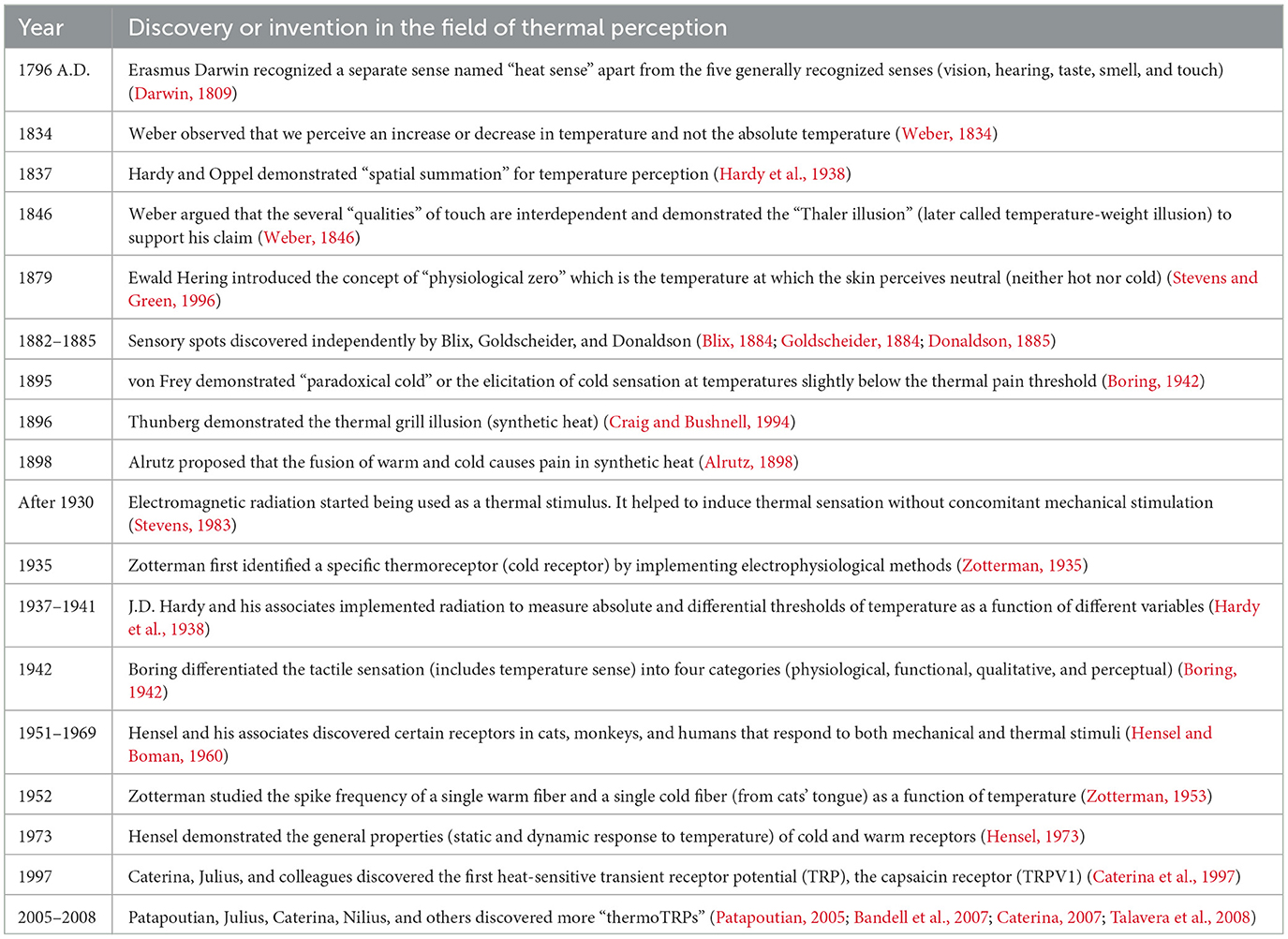
Table 1. Chronology of historical events in thermal perception research.
2.1 Recognition of the thermal senseBefore the mid-nineteenth century, the skin was rarely thought of as an organ with several sensory modalities. The five generally recognized senses were vision, smell, hearing, taste, and touch (Stevens and Green, 1996). In 1796, Erasmus Darwin postulated the presence of more senses in his book “Zoonomia,” in addition to these five senses. Such a sense was the sensation of temperature, which he called a “heat sense” (Darwin, 1809).
In his publication named “Der Tastsinn und das Gemeingefuhl” (“Touch and Common Sensibility”), Weber (1846) first differentiated “touch” into several qualities. He differentiated between the skin's capacity to detect changes in pressure and temperature and its ability to localize sensations on the body. Weber argued that these qualities are interdependent, and to back up his assertions, he employed the “Thaler illusion,” in which a cold silver coin on the forehead seems heavier than a similar coin at a neutral temperature (neither warm nor cold) (Weber, 1846; Stevens and Green, 1996).
According to the German physiologist Ewald Hering, warmth and cold are intertwined with each other in the form of a “single thermal sense” (Boring, 1942). He claimed that warmth and cold are “opponent processes” and the same temperature might feel warm or cold depending on the Nullpunkt, or the “physiological zero,” the temperature that feels neutral (Stevens and Green, 1996).
2.2 Discovery of sensory spotsSensory spots are a key discovery in the domain of thermal perception (Stevens and Green, 1996). A sensory spot is a small area of skin that produces a sensation when stimulated by pain, pressure, or temperature. Between 1882 and 1885, three independent laboratories, one led by Magnus Blix in Sweden, one by Alfred Goldscheider in Germany, and one by Henry Donaldson in America, were engaged in the discovery of these sensory spots (Blix, 1884; Goldscheider, 1884; Donaldson, 1885). Blix observed that electrical stimulation of different sites of the skin can make people feel warm or cold. He mapped the warm and cold sensitive spots using a temperature stimulator made out of a hollow metal cone. Blix's findings were validated and expanded upon by Donaldson. Goldsheider also mapped cold, warm, and pressure sensory spots, concluding that they were all distinct characteristics within the tactile modality. The mappings of the sensory spots revealed that the cold spots outnumber the warm spots by a substantial margin. In addition, the concentration of cold and warm spots varied significantly across the body. The number of cold spots on the lips is significantly higher than on the sole, while the number of warm spots on the fingers is much higher than on the dorsal side of the upper arm (Strughold, 1931).
Spatial summation refers to the phenomenon wherein the perceived magnitude of a sensation increases as the area of skin stimulation becomes broader. Hardy et al. (1938) conducted studies aimed at comprehending the phenomenon of spatial summation in relation to temperature stimuli. Spatial summation will be discussed in more detail later in this paper. The lack of warm spots in some parts of the body does not necessarily indicate insensitivity to warm stimuli. In sites characterized by a small number of warm spots, the application of a stimulus with a greater area of contact (ranging from 1 to 2 cm2) compared to the conventional spot mapping technique (often utilizing a surface area of 1–2 mm2) might induce the sensation of warmth. Therefore, it can be asserted that thermoreceptors and temperature spots are not synonymous terms. It is possible that the number of receptors exceeds the number of spots. A sensation might be produced by the simultaneous activation of several of these receptors (Stevens and Green, 1996). In addition, Maximilian von Frey made the inclusion of pain spots and paradoxical cold spots to the list of sensory spots. Paradoxical cold spots generate cold sensations even at warm temperatures between 45 and 50°C (Boring, 1942). Further technological improvements paved the way for electrophysiological research on thermoreceptors, providing a clearer idea.
2.3 Electrophysiological studies of thermoreceptorsThermoreceptors are sensory units responsible for conveying temperature information from the skin to the brain. They are present in the dermal and epidermal skin layers (Hensel, 1974). The electrophysiological recordings of the individual thermoreceptor fibers helped gain insights regarding the neurophysiology behind temperature sensing (Stevens and Green, 1996). Radiometry advancements enabled the use of electromagnetic radiation as a stimulus for warmth or pain and helped to eliminate the mechanical stimulation that had been present in prior studies (Stevens, 1983). This led to a more effective stimulus for evaluating warmth and pain. Recordings of neural impulses in the cutaneous nerve fibers were done for different species, including human beings.
Different kinds of nerve fibers were found to respond differently. Some nerve fibers responded to temperature alone, whereas others to both temperature and mechanical stimuli. One type of fiber showed an overshoot of spike rate when the skin was heated and a transient inhibition of cooling the skin. On the other hand, certain fibers showed an increase in the rate of impulses when they were cooled and a brief inhibition when they were warmed (Hensel, 1973; Hallin et al., 1982). Further research has aided our understanding of the physiology of the nerve fibers involved in temperature sensing as well as the broader mechanisms at work.
3 Physiology and mechanisms of thermal perceptionThe physiology of temperature perception can be understood from different perspectives. Electrophysiological studies were conducted to observe the thermoreceptors and the afferent fibers involved in thermoreception. This section will go through these aspects in order to better comprehend the thermal illusions that will be discussed in the following sections.
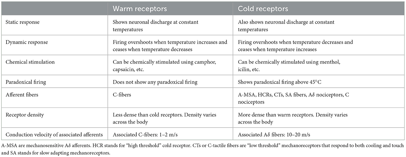
3.1 ThermoreceptorsThe discovery of thermoreceptors and their physiology has helped researchers gain insights into the fundamental processes that govern temperature perception. Thermoreceptors are sensory units that are located beneath the skin throughout the body and are stimulated by temperature changes. In fact, there are different types of receptors for the conduction of warm and cold sensations, namely, “warm” and “cold” receptors, respectively (Stevens, 2013). Thermal information is transported from thermoreceptors to the cortex in the somatosensory area 1 (S1) through the spinothalamic pathway (Milenkovic et al., 2014). Moreover, the warm and cold receptors respond differently to different thermal stimuli (Table 2).
Table 2. Similarities and differences between warm and cold receptors.
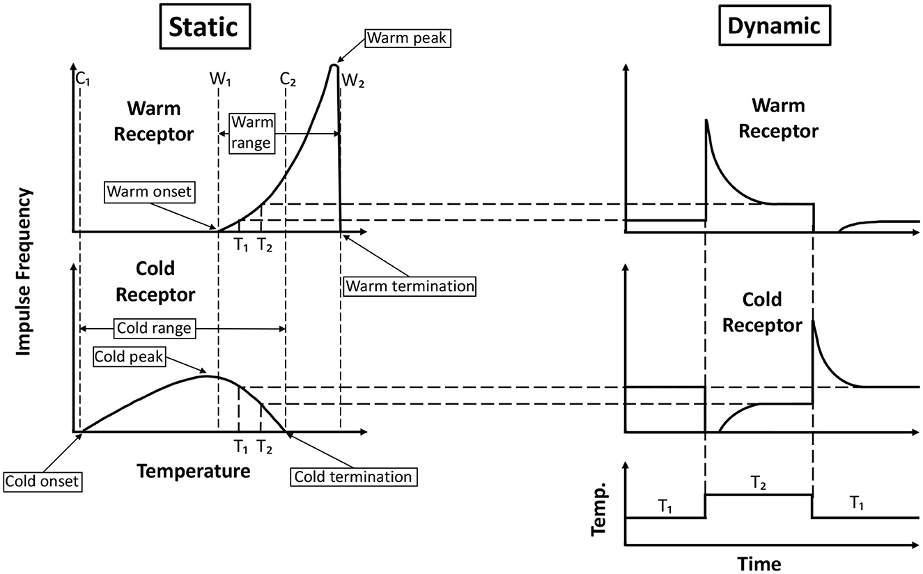
Both the warm and cold receptors are spontaneously firing at normal skin temperatures, and there is no noticeable sensation due to this spontaneous activity. However, when the skin is heated, the warm receptors fire vigorously, and the cold receptors cease the spontaneous firing. Conversely, on cooling, the cold receptors show increased firing while the warm receptors stop their spontaneous firing. Hensel (1973) showed the response of cutaneous single warm and cold receptors present in the cat's nose to constant temperatures (static response) as well as to rapid changes in temperature (dynamic response) (Figure 1). Zotterman (1953) observed that the cold receptors also show a “paradoxical” firing at temperatures above 45°C. As a result, thermoreceptor activity can be conceived of as a combination of the response to a temperature value and the magnitude of change in that value.
Figure 1. Generalized response of cold and warm receptors recorded from cat's nose. The static response is the response at a constant temperature, and the dynamic response is the response when there is a rapid change in temperature. Onset and termination of cold receptor static response are intersected by vertical lines C1 and C2, respectively. Similarly, lines W1 and W2 intersect the onset and termination of the static response of warm receptors. The peak of the cold static response lies very close to the line W1. There is an overlap in the static response range of the warm and cold receptors between W1 and C2. Adapted and redrawn from Hensel (1973).
Cold is perceived much faster than warmth due to the difference in the velocities of the afferent fibers (nerve fibers that carry information from the sensory receptors to the brain) associated with them. The conduction velocity of afferent fibers originating from warm receptors typically falls within the range of 1–2 m/s, but the conduction velocity of afferent fibers originating from cold receptors can be 10–20 m/s (Darian-Smith, 1984). Apart from these warm and cold receptors, there are pain-sensitive nociceptors that activate when a noxious thermal (painfully hot or cold) or mechanical stimulus is applied (Stevens, 2013).
Thermoreceptors can also be stimulated chemically, mimicking the perception of warmth or cold. A number of chemical substances can behave as agonists for temperature perception. For instance, menthol is known to elicit a cold sensation on the skin (Hensel, 1973). Calcium, on the other hand, can produce warmth when applied intracutaneously or intravenously. Calcium and chloroform both have the ability to lower warm thresholds (Schreiner, 1936; Schmidt, 1949). Carbon dioxide and carbonic acid, when applied locally, also produce warmth (Liljestrand and Magnus, 1922; Gollwitzer-Meier, 1937). Certain spices, such as capsaicine, undecylenic acid vanillylamide, and cinnamonylic-acrylic acid piperidide can induce hot as well as pain sensations (Stary, 1925; Sans, 1949). The neuronal activities in the thermoreceptors are mediated by the afferent nerve fibers to the insula cortex (Craig et al., 2000).
3.2 Afferent nerve fibers associated with thermosensationThe majority of the afferent nerve fibers involved in temperature sensation are C fibers and A δ fibers. C fibers are unmyelinated fibers with a lower conduction velocity, whereas the Aδ fibers are thinly myelinated with a higher conduction velocity. Studies on humans have shown that C fibers are activated by cold temperature stimuli in the innocuous range (Campero et al., 2001). Apart from these C and Aδ fibers, there are “high threshold” cold receptors (HCRs) found in monkeys' skin (LaMotte and Thalhammer, 1982). “Low threshold” mechanoreceptors termed C tactile fibers (CTs) respond to rapid cooling as well as light touch (Kumazawa and Perl, 1977). In fact, about half of the slowly adapting mechanoreceptors (SA fibers) like Merkel discs and Ruffini endings respond to cooling stimulus from normal skin temperature to 14.5°C (Hensel and Zotterman, 1951; Duclaux and Kenshalo, 1972; Cahusac and Noyce, 2007). Warm fibers, on the other hand, are C fibers that are insensitive to mechanical stimuli (Darian-Smith et al., 1979). Apart from innocuous warmth and cold, afferent fibers are involved in painful thermal perception.
Both “cold pain” and “heat pain” are mediated by C and Aδ nociceptors (Kumazawa and Perl, 1977; LaMotte and Thalhammer, 1982; Campero et al., 1996; Simone and Kajander, 1997; Schepers and Ringkamp, 2010). “Cold pain” is felt in glabrous (non-hairy) skin at temperatures between 10 and 15°C and in hairy skin at temperatures below 18°C (Davis, 1998; Harrison and Davis, 1999).
An intriguing observation regarding “heat pain” is that it is felt twice and is called a “dual pain sensation” (Campbell and LaMotte, 1983). When noxious heat stimuli were applied to the hairy skin (of the lower arm, dorsum of the hand, and foot), they produced two distinct pain sensations (Campbell and LaMotte, 1983). The first pain was a sharp, pricking sensation that occurred almost 0.4 s after the stimulus. Again, after 1–2 s of the first pain, the second pain was felt as a dull, burning sensation. This dual pain sensation was felt at the distal extremities and tended to merge in the proximal regions of the body. Also, for glabrous skin, like the skin of the palm, the dual pain was not induced (Campbell and Meyer, 1983). This dual pain sensation indicates that two types of fibers are involved in the mediation of “heat pain.” The first pain is mediated by Aδ fibers which have faster conduction velocities, and the second pain is mediated by slower-conducting C fibers (Price et al., 1977; Campbell and LaMotte, 1983).
4 Psychophysics of temperature perceptionPsychophysics is the study of identifying a quantitative link between an external physical stimulus and the conscious perception of the sensation induced by the stimulus (Engen, 1988). Various psychophysical studies were conducted to investigate the underlying mechanisms and unique features of thermosensation. Some properties of thermal sensation, such as the two-point discrimination threshold, thermal detection threshold, thermal adaptation, thermal spatial summation, and thermal localization, are discussed below (Stevens, 2013).
4.1 Two-point discrimination thresholdThe two-point discrimination threshold (TPDT) is often used to measure the spatial acuity of the sensory system. TPDT is the minimum distance between two cutaneous point stimuli at which they are perceived as two separate points instead of a single point. In a recent study by Frahm and Gervasio (2021), TPDT for mechanical and thermal stimuli on the right volar forearm was compared. They observed that the TPDT varied depending on the modality (mechanical or thermal) and the intensity (innocuous or noxious) of the stimuli. The TPDT of a noxious thermal stimulus (66.9 mm) was less compared to that of an innocuous thermal stimulus (80.5 mm). On the contrary, the TPDT of a noxious mechanical stimulus (47.1 mm) was found to be greater than the innocuous one (34.7 mm) (Frahm and Gervasio, 2021).
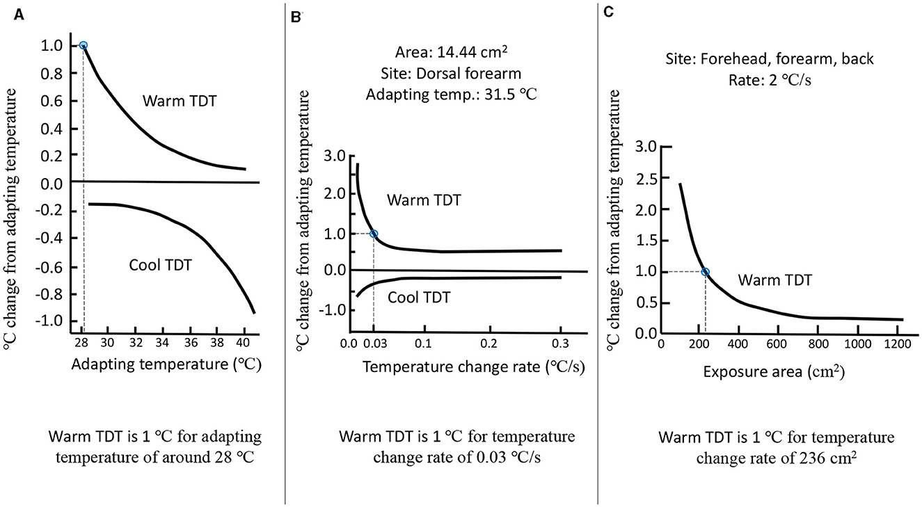
4.2 Thermal detection thresholdThe thermal detection threshold (TDT) is the lowest temperature difference that can be perceived during an increase or decrease from the baseline temperature (Kappers and Plaisier, 2019). Studies show that the TDT depends on at least four parameters (Lee et al., 1998; Jones and Berris, 2002). The four parameters are:
1. The temperature of the skin stimulated.
2. The rate of change of temperature.
3. The area of the skin stimulated.
4. The site of the skin stimulated.
With the increase in the adapted skin temperature, the TDT for warm sensation decreases, whereas the TDT for cool sensation increases (Figure 2A). At a skin temperature of 34°C, the warm TDT was measured to be 0.3°C, while the cool TDT was found to be 0.2°C. These measurements were obtained using a thermal stimulus of a certain area and a controlled rate of temperature change (Lee et al., 1998). For a fixed skin temperature, the thermal TDT depends on the rate of temperature change of the stimulus (Figure 2B). A higher rate of temperature change results in a lower warm TDT and a higher cool TDT. If the temperature is within the neutral range (30–36°C), then an increase of 5–6°C goes unnoticed by human subjects at a rate of 0.5°C/min. On the other hand, even a small increase in temperature can be detected at a much faster rate of 0.1°C/s (Kenshalo et al., 1968; Kenshalo, 1978). The area of the skin that is thermally stimulated also influences the TDT for thermosensation (Figure 2C). As the skin exposure area increases, the warm TDT decreases, whereas the cool TDT increases (Kenshalo et al., 1967; Stevens and Marks, 1971). Jamal et al. (1985) observed that the warm and cold thresholds were much higher for the ankle compared to the wrist, forearm, or thigh. In another study of thermosensitivity, 49 sites on the hand and 54 on the foot were mapped, and it was found that different regions in the foot can vary in thermosensitivity by a factor of five. Also, glabrous skin was found to be less thermosensitive than hairy skin, which is the opposite for tactile and pain sensitivity (Filingeri et al., 2018). Moreover, thermosensitivity depends on the climate of the place where the person is from (Lee et al., 2010). Inhabitants of tropical climate zones have a reduced sensitivity to detecting warmth. Thermal sensitivity is also influenced by age. The thermal detection thresholds for cold and warm were investigated by Stevens and Choo (1998) in a study with a sample of 60 persons ranging in age from 18 to 88 years. The study revealed a decrease in thermal sensitivity with advancing age, particularly in the peripheral regions, such as the foot.
Figure 2. (A) Thermal detection threshold as a function of adapting skin temperature. (B) Thermal detection threshold as a function of rate of change of temperature. (C) Thermal detection threshold as a function of an area of the thermal stimulation. Adapted and redrawn from Lee et al. (1998).
Several psychophysical studies focused on how the knowledge of thermal detection can be applied to practical experiences. A research investigation was undertaken to examine temperature detection in various regions of the human body where a portable electronic device could potentially be held, including the thenar, finger, forearm, and upper arm (Wilson et al., 2011). Results showed that it was easier to detect the temperature change when the thermal intensity (the increase or decrease in temperature from the baseline) was higher. Moreover, cooling was detected much faster than warming (Wilson et al., 2011). In another study by Halvey et al. (2011), they implemented different types of textiles (nylon and cotton) to investigate the perceptual effect when they were placed between the skin (of the thenar, thigh, and waist) and a thermal stimulus. Participants reported a more comfortable sensation when textiles were used between the stimulus and their skin. The detection performance improved for thermal stimuli of higher intensity and a higher rate of change of temperature. Thermal detection also depended on the type of the textile, and it performed better for nylon than for cotton (Halvey et al., 2011). Ketna and Leelanupab (2017) conducted experiments to assess the perceptual efficacy of different types of stimulation (auditory, vibratory, and thermal) in a challenging environment characterized by high levels of noise and vibration. The study revealed that thermal stimulation exhibited the highest level of detectability in a noisy and bumpy environment. Furthermore, the outcomes were notably enhanced when multimodal stimulation was employed (Ketna and Leelanupab, 2017).
4.3 Thermal adaptationThermal adaptation is the ability of the thermoreceptors beneath the skin to adjust to a temperature stimulus. It is the gradual reduction of responsiveness to thermal stimuli over time (Kenshalo and Scott, 1966). When hands are dipped in warm water, the sensation of warmth gradually fades away, and after a while, the same water at the same temperature might feel cold (as observed by John Locke in the 17th century). Adaptation can manipulate the “physiological zero” of a person by shifting it to a lower or higher temperature (Stevens, 2013). Kenshalo and Scott (1966) conducted a study in which the participants were asked to sit in an air-conditioned room for 20 min followed by a task in which the participants had to adjust the temperature of a Peltier element placed on their forearm so that it was just detected as warm or cold. Several such trials were conducted over a period of 40 min during which the difference between the detection temperatures for warm and cold was noted. During this time period, there was an apparent rise in the difference between detection temperatures, which ranged from ~1°C to around 4°C. The reason behind this increase was thermal adaptation (Kenshalo and Scott, 1966).
4.4 Thermal spatial summationThe term “spatial summation” refers to the increase in perceptual or neurophysiological response when there is an increase in the area of stimulation (Stevens, 2013). It has been observed that thermal thresholds remain constant if the area of stimulus is doubled and the intensity of stimulus is halved (Jones, 2009). However, this phenomenon breakdown is usually near the pain threshold (Paricio-Montesinos et al., 2020). Studies were conducted to investigate spatial summation in different body regions (upper arm, forearm, back, chest, shoulder, and calf). It was observed that the perceived temperature was a power function of the intensity of the stimulus. The power function's exponent depends on the size of the area being stimulated and is smaller for larger areas (Marks, 1971; Stevens and Marks, 1971; Stevens et al., 1974). In another study, it was shown that thermal spatial summation can also happen for non-adjacent areas (areas that are far from each other on the body) of stimulation. In fact, thermal spatial summation can happen over the two arms by stimulating either one forearm or both forearms simultaneously (Rózsa and Kenshalo, 1977).
4.5 Thermal localizationPsychophysical experiments were conducted to investigate the capacity for localizing thermal perception. In a research study, Taus et al. (1975) administered radiant thermal stimuli to the participants' forearms and instructed them to discern the relative proximity of the radiant area to either the wrist or the elbow. The study findings indicate that there was a noticeable decline in performance when the thermal stimulus was of reduced intensity and positioned in closer proximity to the mid-line, which is the midpoint between the wrist and elbow. Conversely, the performance exhibited enhancement as the intensity increased and the distance from the mid-line increased (Taus et al., 1975).
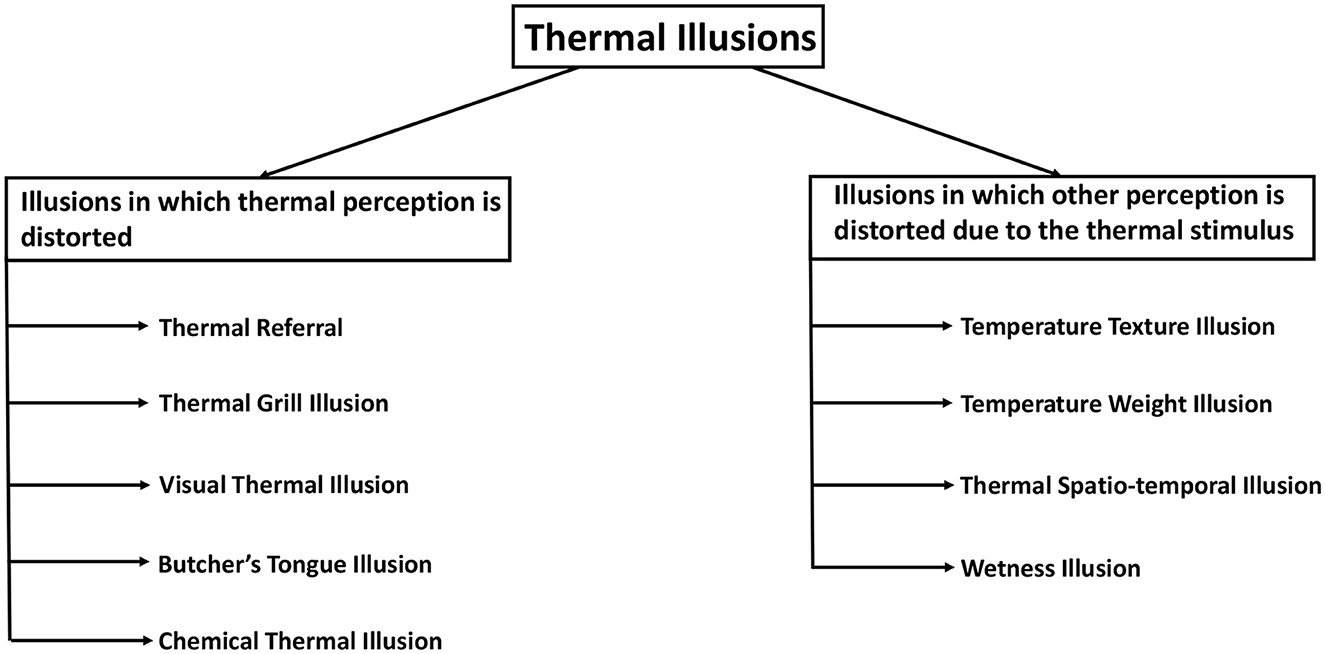
5 Classification of thermal illusionsAs sensory modality research progressed, more and more studies uncovered intriguing temperature perception phenomena in humans. Many of these phenomena present an illusory perception and can be called thermal illusions. An attempt to classify these illusions has been made in this section. Such categorization can aid in identifying the similarities and differences in the nature and mechanisms of thermal illusions. To introduce the breadth of thermal illusions two categories are considered (Figure 3):
Figure 3. Classification of thermal illusions based on the type of sensation getting distorted. The illusions on the left are those in which thermal perception is influenced, whereas the illusions on the right are those in which the thermal stimulus influences another perceptual modality.
1. Illusions in which the thermal perception is distorted.
2. Illusions in which the thermal stimulus distorts other sensory perceptions.
In the first category of thermal illusions, there is a discrepancy between the thermal perception experienced by the subject and the actual thermal stimulus that is presented to the subject. For instance, in Thermal Referral, a person can feel a thermal sensation in a region of the body where no thermal stimulus is present (Green, 1977b). In another illusion of the first category, an intense thermal perception can result from a considerably less intense thermal stimulus. The thermal grill illusion causes a person to experience a burning pain due to interlaced cold and warm stimuli that are individually not painful (Thunberg, 1896). Also, the thermal perception of a person can be manipulated by a stimulus to the other sense modalities. In the context of visual-thermal illusions, it is observed that the visual attributes of an object, such as its color, exert an influence on the corresponding thermal information of the object (Ho et al., 2014). Another similar example is the Butcher's tongue illusion, in which a dummy tongue is made to feel like someone's own using touch sensations, and then a visual-thermal stimulus on that tongue is felt in the real tongue by the subjects (Michel et al., 2014). Furthermore, the phenomenon of Chemical Thermal Illusion enables individuals to perceive sensations of warmth or coldness even in the absence of any actual thermal stimuli (Brooks et al., 2020).
The second category encompasses a collection of illusions wherein the thermal stimulus induces a discrepancy in another sensory modality. In the Temperature texture illusion, the skin temperature causes an alteration of the roughness perception of an object (Green et al., 1979). The perception of weight or force is distorted by the Temperature weight illusion (Weber, 1846). The perception of wetness is induced using a cold dry stimulus in Wetness illusion (Shibahara and Sato, 2016). In fact, temperature perception can also get intertwined with time perception or the ability to localize a stimulus. In the Thermal spatio-temporal illusion, the spatial and temporal properties of thermal stimulus cause distortion in the localization of the stimulus (Trojan et al., 2006; Singhal and Jones, 2016b). Furthermore, thermal illusions have been categorized in different ways with the aim of developing a thermal illusion taxonomy.
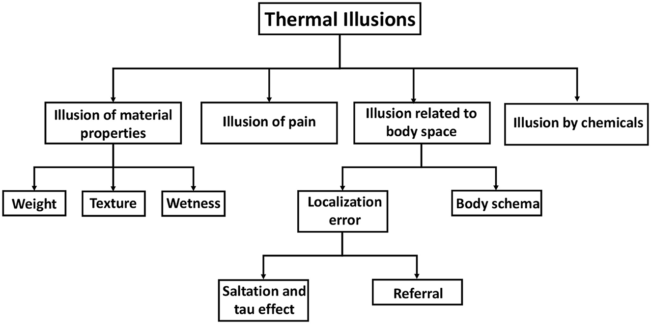
The classification of tactile illusions has been done by Lederman and Jones (2011), as summarized and taxonomically represented by Patel et al. (2019). The taxonomy for tactile illusions has served as a source of motivation and reference for the development of our taxonomy for thermal illusions. Thermal illusions are grouped into four categories or groups (Figure 4). One group involves those thermal illusions which influence the material properties of an object. Temperature-weight illusion, temperature-texture illusion, and wetness illusion fall under this category. The thermal-grill illusion is, however, kept in a separate group named “Illusion of pain” since it is associated with a noxious perception of temperature. The third group constitutes those thermal illusions that are related to the perception of body space by the individual. For instance, in the thermal spatio-temporal illusion, the ability of the subject to localize thermal sensations is distorted. Similarly, in thermal referral, a thermal sensation is perceived in a location where no stimulus is present. Again, the visual-thermal illusion and the Butcher's tongue illusion involve body ownership of an out-of-body object. Hence, they are classified under “Body Schema”. The chemical thermal illusion belongs within the fourth group, specifically labeled as “Illusion by chemicals.” A thorough review of each illusion and its corresponding psychophysical research has been undertaken. We have also endeavored to explore the theoretical foundations underlying some illusions. The objective of these deliberations is to obtain a comprehensive understanding of the research conducted on thermal illusions, with the aim of devising strategies for their application in thermal displays.
Figure 4. Taxonomy of thermal illusion. Thermal illusions are broadly classified into four types: illusions of material properties, illusions of pain, illusions by chemicals, and illusions related to body space. Material properties include the weight and texture of an object that is touched. Body space illusions include illusions related to body schema as well as errors in localizing thermal stimulus.
6 Illusion of material propertiesThe temperature stimulus provided to the skin impacts the perception of an object's material properties (Lederman and Jones, 2011). The illusions in this section can be used as a source of ideas for making haptic devices that show material properties.
6.1 WeightPerception of the weight of an object can be influenced by many properties which are not normally considered to be the sensory information signaling weight. These properties include shape, volume, surface texture, density, and temperature (Jones, 1986). However, since in this review we are only focusing on thermal illusions, discussion of properties other than temperature is beyond the scope.
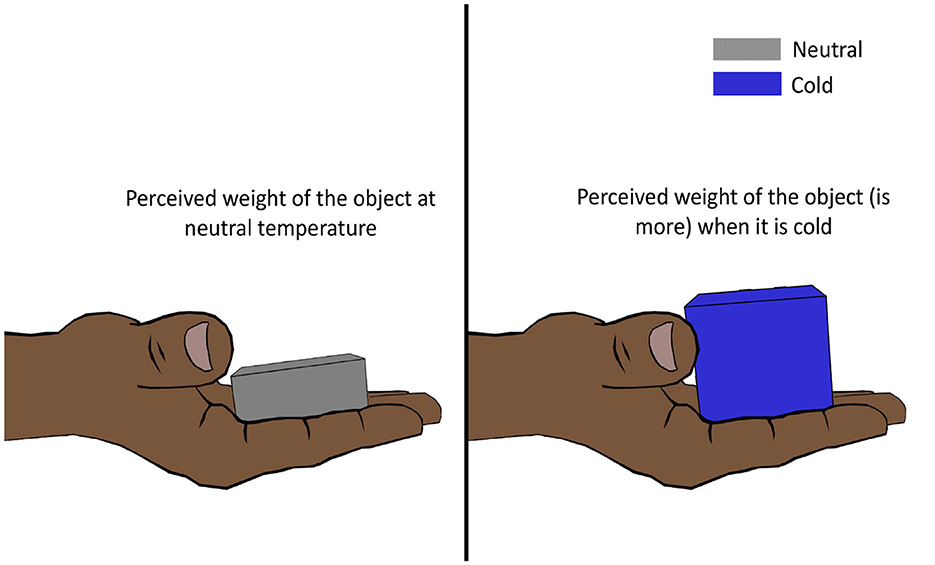
6.1.1 Temperature weight illusionWeber (1846) discovered that placing a cold German dollar (thaler) on the forehead feels almost as heavy as placing two warmer identical dollars on top of the other on the forehead. This led him to conclude that cooling strengthens and warming lightens the pressure sensation on the skin. This then came to be known as Weber's phenomenon, the silver thaler illusion, or more commonly, the temperature weight illusion (TWI) (Weber, 1846; Stevens and Green, 1978; Weber and Ross, 1978). However, later, this theory was proved to be only partially correct by Stevens and Hooper (1982). Stevens and Hooper (1982) hired subjects to estimate the perceived heaviness of metallic stimulators of different masses and temperatures (cold, warm, and neutral), and they found that cold objects (25°C) felt significantly heavier (Figure 5), while warm objects (38°C) felt slightly heavier than neutral objects (33°C). The illusion was shown to be evident in different body regions (hand, arm, abdomen, back, and thigh) as well. Where cold-intensified heaviness was perceived across multiple body sites, warm-intensified heaviness was perceived in some regions (e.g., forearm) and not in others (e.g., forehead) (Stevens, 1979).
Figure 5. Temperature weight illusion. Lowering the temperature of an object increases its perceived weight. For an object at a higher temperature, the illusion is not perceived as strongly.
Controlled force and temperature stimuli were employed in a more recent study to re-evaluate the phenomenon and better comprehend the relationship between the tactile and thermal systems (Dunn et al., 2017). The study involved the application of two types of stimuli: a cooled tactile stimulus, where force was exerted together with a cooling stimulus that reduced the temperature from 32 to 28°C, and a tactile-only stimulus, where force was delivered at a thermally neutral temperature of 32°C. Almost all the participants reported the cooled tactile stimulus to be heavier than the tactile-only stimulus, but the effect is greater when the cooled tactile stimulus is applied after the tactile-only stimulus. In a subsequent experiment, the myelinated fibers of the ulnar nerve of the subjects were blocked (known as compression block) such that there was a loss of vibration (Aβ fibers blocked) and cold sensibility (Aδ fibers blocked), followed by a test where the same load was applied in combination with the cold and neutral temperature (Dunn et al., 2017). In the compression block condition, there was a significant reduction in the incidence of the TWI phenomenon. It was concluded that the TWI is more robust when all the nerve fibers are intact and that both the myelinated and unmyelinated fibers contribute to the phenomenon (Dunn et al., 2017).
Studies were also conducted to investigate the relationship between temperature and actively generated forces. Galie and Jones (2010) conducted an experiment to test whether the temperature of a surface has any effect on the perceived magnitude of the force applied by the finger on that surface. Results showed no such relationship between temperature and the perceived magnitude of actively generated force (Galie and Jones, 2010). However, in a recent study by Kuhtz-Buschbeck and Hagenkamp (2020), the effect of the TWI was observed in a grip-lift experiment where the subject lifted the test objects at varying temperatures (cold, neutral, and warm) from the palm of the non-dominant hand using the dominant hand. Recordings of the grip and lift forces revealed that the subjects applied more force (10% higher) when lifting a cold (18°C), heavy (700 g) object compared to a neutral object of the same weight (Kuhtz-Buschbeck and Hagenkamp, 2020).
Interestingly, the temperature of a surface in contact with the skin can generate the illusion of a push or pull. In a study by Watanabe and Kajimoto (2015), the participants were asked if they perceived any force on touching temperature-changing thermal elements. They reported an illusory sensation of pressure or push when the temperature of the display increased rapidly (27–37°C). Conversely, a pulling sensation was reported with a fast temperature drop (37–27°C) of the thermal display (Watanabe and Kajimoto, 2015). However, these psychophysical investigations are inadequate to describe the illusion's mechanism. The role of mechanoreceptors during temperature change can help explain the occurrence of TWI.
There are many slow-adapting (SA) mechanoreceptors that respond to both skin deformation and skin cooling and hence can be responsible for the TWI (Johnson et al., 1973). It is hypothesized that the activation of such receptive afferent fibers when holding a cold object results in the thermal intensification effect, causing the TWI (Lederman and Jones, 2011).
6.2 TextureSkin temperature has been found in several studies to affect how an object's texture is perceived (Green, 1977a; Bolanowski et al., 1988; Zhang et al., 2009; Jones and Singhal, 2018). Understanding the relationship between skin temperature and texture perception has become more essential with the introduction of multi-dimensional tactile displays.
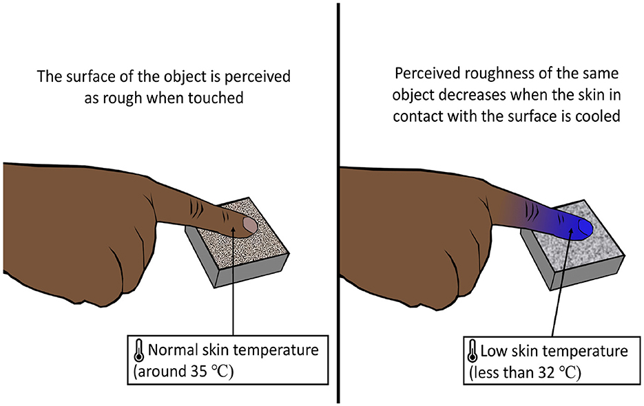
6.2.1 Temperature texture illusionThe temperature of the skin has an effect on the roughness perception of an object in contact with the skin (Green et al., 1979). The apparent roughness declined as the skin temperature fell below 32°C (Figure 6). On the contrary, apparent roughness tended to increase with the increase in skin temperature although the effect was more dominant for cooling (Green et al., 1979). The phenomenon of alteration of texture perception of the surface of an object caused by a change in skin temperature can be called the temperature texture illusion (TTI).
Figure 6. Temperature texture illusion. Lowering the skin's temperature reduces the skin's ability to perceive a rough surface, influencing texture perception.
Temperature also affects the vibrotactile sensitivity of the skin. The sensitivity of the skin to vibratory stimuli of varying frequencies decreased when warmed or cooled. Cooling, on the other hand, had a greater effect than warming on reducing sensitivity (Weitz, 1941; Green, 1977a; Bolanowski and Verrillo, 1982). Hence, similar to the temperature weight illusion, the TTI is also a result of the dependence of mechanoreceptors on temperature.
6.3 WetnessResearch suggests the perception of wetness is achieved by the integration of both tactile and thermal cues, as there are no specialized receptors that are dedicated to the sensation of wetness (Clark and Edholm, 1985; Filingeri et al., 2014b; Filingeri and Ackerley, 2017). The significance of the feeling of wetness lies in its capacity to enable humans to mitigate potential health hazards by avoiding unfavorable environmental conditions. For instance, the discomfort experienced when wearing damp clothing serves as a signal to dispose of the wet garment and prevent getting sick (Chau et al., 2018). This section explores the phenomenon of illusory perception of wetness.
6.3.1 Wetness illusionThe wetness illusion refers to a phenomenon in which the perception of wetness is erroneously inferred by an individual due to the combined influence of thermal and tactile inputs. The sense of wetness is more sensitive to dynamic touch compared to static touch (Bergmann et al., 2012; Filingeri et al., 2014b). Dynamic touch refers to a tactile interaction when a contact surface undergoes relative movement over the surface of an object. In contrast, static touch involves a stationary contact area that does not exhibit any movement. The study revealed that a decrease in temperature is associated with an increase in the perception of wetness during static touch. However, in the case of dynamic touch, a decrease in temperature did not result in an increase in the perception of wetness (Filingeri et al., 2014b). The study conducted by Shibahara and Sato (2019) yielded similar findings, indicating that the illusion of wetness is more effectively induced through static touch rather than dynamic touch.
Wetness perception can be evoked using a dry and cold stimulus (Daanen, 2009; Bergmann Tiest et al., 2012; Filingeri et al., 2013, 2014a,c). Shibahara and Sato (2016) augmented the sensation of a wet cloth by controlling the temperature and softness of a dry cloth. The experiment demonstrated that the perception of wetness in a cloth can be replicated by manipulating two factors: reducing the temperature and enhancing the material's softness or compressibility. In a recent study conducted by Han et al. (2020), a prototype called Mouill was developed with the aim of inducing the illusion of wetness on the user's fingers during the manipulation of hard and soft items. A demonstration was given of a virtual reality application utilizing the device, showcasing the simulation of various things such as an ice cube, damp sponge, and chilled cola bottle.
7 Illusion of painThis section is dedicated to the illusory perception of thermal pain. Currently, the thermal grill illusion is the only known phenomenon to induce the noxious perception of temperature using innocuous temperature stimuli.
7.1 Thermal grill illusionIn the 19th century, Thunberg (1896) and Alrutz (1898) independently discovered that interlaced warm and cold stimuli on the skin elicit a paradoxical sensation of burning pain. It was referred to as “synthetic heat” or the “thermal grill illusion” (Figure 7) (Fardo et al., 2020). Although the existence of the illusion was questioned by Jenkins (1938), influential research (Green, 1977a,b, 2002; Craig and Bushnell, 1994; Craig et al., 1998; Bouhassira et al., 2005) eradicated all the doubts, and thermal grill illusion (TGI) again became the object of study among researchers. The term “synthetic heat” was mostly used while investigating illusory heat without pain (Burnett and Dallenbach, 1928; Green, 1977b, 2002). On the other hand, the term “thermal grill illusion” denotes a thermo-nociceptive prickling sensation elicited by the grill of alternating warm and cold bars, known as the thermal grill. In studies, similar apparatus, such as alternating cold and warm spiral tubes (Thunberg, 1896; Bach et al., 2011) or alternating warm and cold thermodes (Defrin et al., 2002; Kammers et al., 2010; Marotta et al., 2015; Fardo et al., 2018), were used to elicit the illusory pain.
Figure 7. Thermal grill illusion. When an innocuous warm or cool stimulus is applied to the palm, a non-painful warmth or cold sensation is felt, respectively. However, when the same innocuous warm and cold stimuli are applied in an interlaced manner, a painful burning sensation is perceived.
Though earlier studies showed that thermal grill illusion (TGI) can evoke a wide range of sensations (Burnett and Dallenbach, 1928), more recent studies suggest that TGI evokes mainly pain (Craig and Bushnell, 1994; Bouhassira et al., 2005; Kern et al., 2008a; Posada-Quintero et al., 2020). Several qualitative and quantitative approaches have been taken to understand the perception felt by the TGI. Subjects were asked to select from subjective descriptors of the illusion (e.g., pricking, cutting, stinging, burning, scalding, freezing, etc.) (Li et al., 2009; Bach et al., 2011), rate the illusion on verbal-numerical scales (e.g., verbally rate the pain between 0 and 100) (Scheuren et al., 2014) and visual analog scales (e.g., rate the pain on a visual scale of 100 mm) (Craig and Bushnell, 1994; Leung et al., 2005; Kern et al., 2008a,b; Adam et al., 2014), and also perform temperature matching tasks (Leung et al., 2005; Kammers et al., 2010; Marotta et al., 2015; Fardo et al., 2018; Ferrè et al., 2018). Factors such as genetics, gender, and psychological disorders were also found to impact the perceived intensity of TGI (Lindstedt et al., 2011; Averbeck et al., 2017). In the study by Averbeck et al. (2017), women reported a lower TGI-pain threshold than men. Patients with psychological disorders such as borderline personality disorder (BPD) or major depressive disorder (MDD) experienced less TGI intensity than healthy subjects (Boettger et al., 2013; Bekrater-Bodmann et al., 2015). Despite the fact that the neurophysiological mechanism underlying the phenomenon remains unknown, two significant theories have emerged.
“Addition or convergence theory” and “disinhibition or unmasking theory” are the two popular theories of TGI. The previous assumption in the late 19th century, known as the “addition theory,” was that concurrent warm and cold activation fuses into a sensation of heat. Craig and Bushnell (1994) proposed the “disinhibition theory,” according to which the TGI results from the disinhibition of C-polymodal nociceptors (heat-pinch-cold or HPC neurons) by the COOL neurons (i.e., neurons activated by mild cold temperatures) in the spinal cord (Craig and Bushnell, 1994; Craig et al., 1996, 1998). Both the theories are based on Johannes Müller's “law of specific nerve energies,” which postulates that for every sensory quality, there exists a distinct afferent pathway that receives signals from specific peripheral receptors and transmits them to the associated cortical targets in the brain, which finally determines the perceptual quality of the sensation (Norrsell et al., 1999; Fardo et al., 2020). Thus, for each sensation of heat, cold, or pain, there is a dedicated pathway or “labeled line” to transmit signals from the periphery to the brain.
The “addition theory” suggests that a polymodal pathway, activated by both warm and cold sensory inputs, encodes the linear summation of the inputs, resulting in the TGI (Green, 2002; Bouhassira et al., 2005). The greater the difference between the temperatures of the warm and cold sensory stimuli, the greater the perception of the synthetic heat caused by neural summation (Burnett and Dallenbach, 1927). Burnett and Dallenbach (1928) and Gritman and Dallenbach (1929) developed mathematical models based on the assumption that the inputs from both warm and cold temperatures activate polymodal pathways, and then the summation of the inputs causes increased firing rates, mimicking that of painful stimulation. The polymodal pathways are proposed to be second-order wide-dynamic range (WDR) neurons in the dorsal horn of the spinal cord (Green, 2002; Bouhassira et al., 2005). These WDR neurons, which respond to mechanical and thermal stimulation, are suggested to be involved in the painful perception of both noxious stimulation and that of thermal grill (Mendell, 1966; Maixner et al., 1986; Hao et al., 1992; Coghill et al., 1993; Khasabov et al., 2001). However, a more accepted theory of TGI is that of the “disinhibition theory.”
The “disinhibition theory” suggests that there are two separate steps involved in the painful perception of TGI (Craig and Bushnell, 1994; Craig et al., 1998; Craig, 2003). First, the warm stimulation causes the inhibition of COOL neurons in the dorsal horn of the spinal cord. The COOL neurons respond to temperatures below 27°C and are assumed to inhibit pain at the thalamo-cortical level. Second, due to the reduced inhibition of the COOL neurons, the pain-sensitive C-polymodal nociceptors (HPC neurons) get disinhibited. Hence, the “warm inhibits cold” mechanism results in the reduction of the “cold inhibits pain” mechanism, which finally leads to “disinhibition of pain.” These assumptions of the mechanism of “warm inhibiting cold” and “cold inhibiting pain” are based respectively on the electro-physiological studies conducted by Craig and Bushnell (1994) and the observations of nerve blocks altering cold, warm, and pain perception (Mackenzie et al., 1975; LaMotte and Thalhammer, 1982; Fruhstorfer, 1984; Wahren et al., 1989; Yarnitsky and Ochoa, 1990). A mathematical model of TGI based on the “disinhibition” theory has been proposed by Karmakar et al. (2023). Both “addition” and “disinhibition” theories are based on the recruitment of a specific type of neuron (WDR or HPC) for painful perception. Nevertheless, Fardo et al. (2020) recently proposed an alternative hypothesis based on the theories of population coding.
Population coding models are widely accepted in vision neuroscience and are based on the pattern theory of color vision and the across-fiber pattern approach (Erickson, 1973). The hypothesis proposed for TGI was inspired by the gate control theory (Melzack and Wall, 1962, 1965). It supported the view that perception relies upon spatio-temporal patterns of activity in the central nervous system, distributed across nociceptive and non-nociceptive pathways (Melzack and Wall, 1962, 1965). The main concept of population coding was that perception is probabilistically dependent on the neural response of a distinct population of neurons at different levels of neuraxis (Averbeck et al., 2006; Quiroga and Panzeri, 2009; Panzeri et al., 2015). Hence, Fardo et al. (2020) hypothesized TGI as a consequence of a certain neuronal pattern which is interpreted as a burning pain in the brain. The mechanism behind TGI has been a mystery for decades, and researchers are still trying to unravel it. Understanding TGI might also help researchers with studying chronic pain perception and pain management (Shin and Chang, 2021). Attempts to incorporate the TGI into thermal displays have been made (discussed in later sections).
8 Illusions related to body spaceThis section discusses illusions related to the spatial representation of the body. The category is again divided into two sub-categories, namely, localization error and body schema. A localization error is one in which there is a distortion in the perception of the stimulus location. Body schema is the awareness of the relative positions of distinct body parts in external space.
8.1 Localization errorThe error in localization can happen either due to the non-uniformity of the stimuli as in Thermal Referral or due to the influence of other perceptual information as in Thermal Spatio-temporal Illusion. Each of the two illusions has been discussed below.
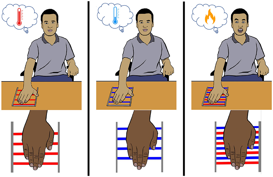
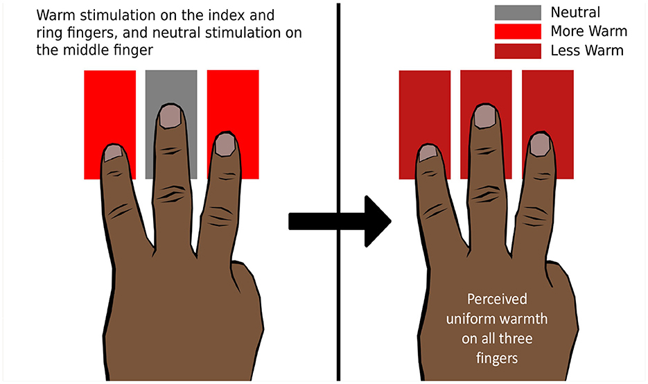
8.1.1 Referral: thermal referralGreen (1977b) demonstrated that when the index and annular fingers are thermally stimulated by hot or cold temperatures while the middle finger is kept on a thermally neutral stimulator, the middle finger also perceives hot or cold temperatures, respectively. This illusion is called “thermal referral” (TR) since the thermal sensation at the outer fingers is referred to the middle finger (Figure 8). However, the illusion disappears when the middle finger is lifted or kept just above the stimulator (Green, 1977b). Hence, it is necessary to stimulate the middle finger with a tactile sensation for the phenomenon to occur. TR can also be elicited using two fingers by giving tactile stimulation to one and thermal-tactile stimulation to the other. TR was elicited in other areas of the body as well, including the forearm (Green, 1977b).
Figure 8. Thermal referral. The ring and index fingers are presented with a warm thermo-tactile stimulus, and the middle finger is presented with a neutral tactile stimulus. All three fingers perceive uniform warmth, which is less intense than the physical intensity of the warm stimulus.
In a study by Ho et al. (2011), it was observed that the sensation of cold or hot is uniform across three fingers during TR, but the perceived intensity on the outer fingers is lower than the physical intensity applied to the outer fingers (Ho et al., 2011). It is proposed that the TR phenomenon is mediated by two stages. First, the thermal perceptions across multiple stimulated areas are homogenized to determine an overall intensity perception. In the second stage, this resulting intensity is referred to or localized to different regions of the body based on the tactile stimulation (Ho et al., 2011). Ho et al. (2010) also investigated how thermal and tactile modalities coordinate to process the localization information.
The strength of mislocalization in TR depends upon the somatotopic distance or the distance between the sites represented in the cortical topography. The strength of the illusion is reduced as the somatotopic distance increases, but the spatiotopic distance, or the distance between the sites on the body, has no effect (Ho et al., 2010).
Comments (0)