
Remember me
 Introduction
Split cord malformation (SCM) is a rare congenital anomaly within the spectrum of occult spinal dysraphisms (OSDs), involving longitudinal splitting of the spinal cord by a fibroosseous or fibrous septum. It is frequently associated with tethered cord syndrome, vertebral anomalies, lipomas, and dorsal dermal sinuses. Pang et al. classified SCM into Type I (diastematomyelia) and Type II (diplomyelia) based on the septal composition and dural sac configuration. Although SCM accounts for ~5% of congenital spinal defects, antenatal diagnosis is rare. We present a mid-trimester prenatal case of SCM with tethered cord, posterior lipoma, vertebral anomalies, dorsal dermal sinus, and unilateral club foot, diagnosed on ultrasound and confirmed postmortem. Multiplanar high-resolution ultrasound revealed a widened spinal canal, segmental cord splitting, low-lying conus, and a fat-intense lesion abutting the lamina and extending into the subcutaneous plane. Postmortem imaging confirmed these findings, including dural calcification and vertebral anomalies.
Understanding normal fetal spine anatomy is essential for identifying subtle deviations. In the mid-trimester, the spine appears as two echogenic lines in sagittal views, representing anterior and posterior ossific centres. The spinal cord is seen as a hypoechoic structure with a central echogenic canal, with the conus typically terminating at L2–L3 ([Fig. 1a and b]). Axial views show one anterior and two posterior ossific centres enclosing the vertebral canal, which contains the cord as a hypoechoic circle with a central dot ([Fig. 1c]). In ventral coronal sections, anterior centres appear ventrally, while posterior centres form two parallel rows converging toward the sacral tip ([Fig. 1d]).
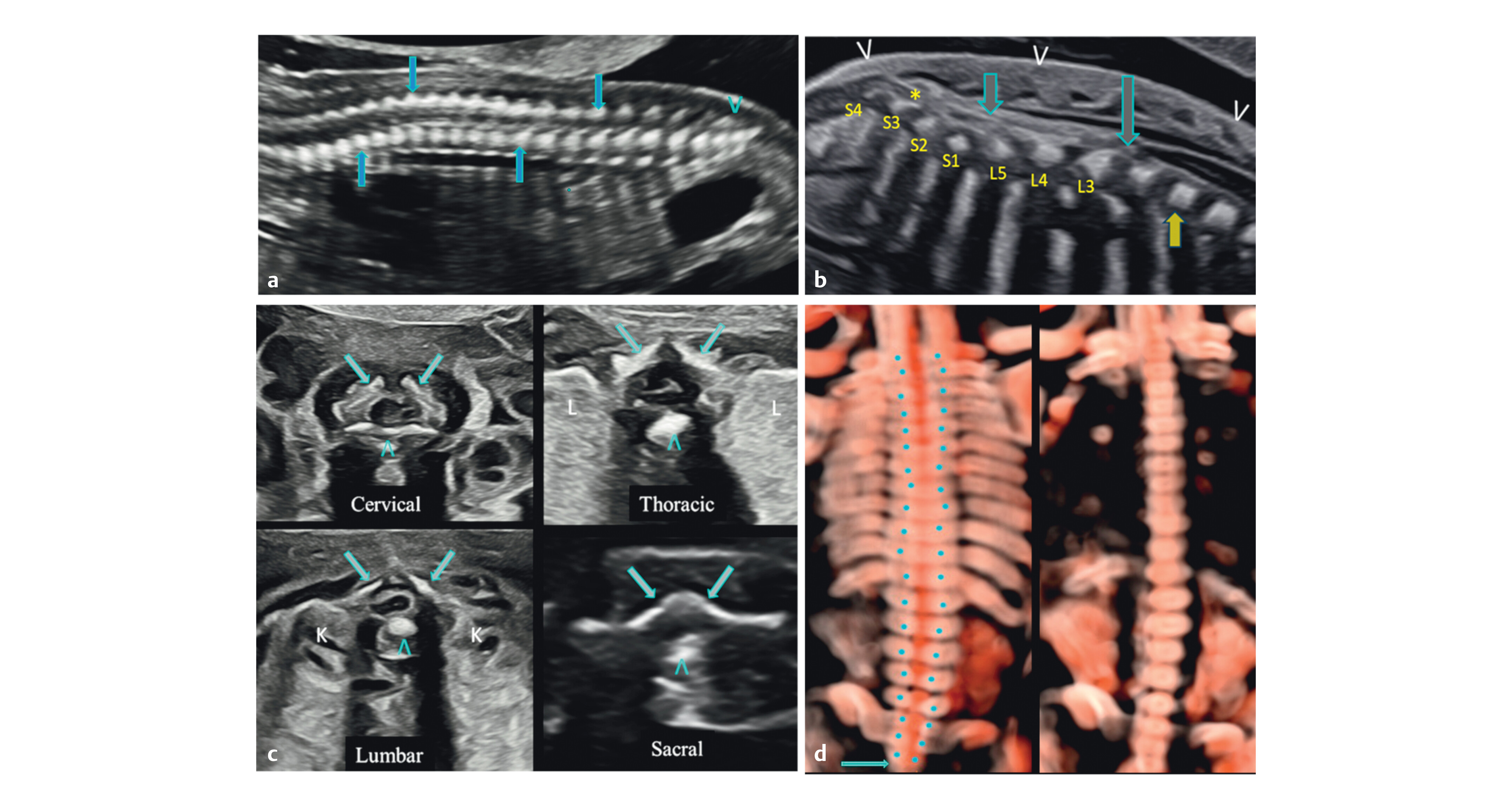 Fig. 1 Mid trimester sections of the normal spine: (a) A
paramedian sagittal section shows the posterior ossific centres (downward
arrow), anterior ossific centres (upward arrow), and two rows converging to
form the sacral taper with dorsal uptilt (arrowhead). (b) A
midsagittal section of the normal lumbosacral spine—only the anterior
ossific centre row is seen (solid arrows), conus medullaris (long arrow),
cauda equina (short arrow), and filum terminale (*). Conus medullaris is at
the lower border of L3. Counting is done from S4 cephalad or from T12
caudal. Intact skin layers seen over the entire length of the spine
(arrowheads). (c) Axial sections of the cervical, thoracic, lumbar
and sacral spine—two posterior ossific centres (arrows), one anterior
ossific centre (arrowhead), lungs (L), kidneys (K), and spinal cord is seen
in the vertebral canal as an ovoid hypoechoic structure fillied with CSF and
a central echogenic dot or an oval structure corresponding to the cord,
except at the sacral level where the cord with cauda equina is completely
echogenic. (d) Coronal sections, the left posterior section showing
the posterior ossific centres forming two parallel rows (rail road
appearance) converging at the sacral tip (arrow) and the right anterior
section showing the anterior ossific centres as a single row.Publication History
Fig. 1 Mid trimester sections of the normal spine: (a) A
paramedian sagittal section shows the posterior ossific centres (downward
arrow), anterior ossific centres (upward arrow), and two rows converging to
form the sacral taper with dorsal uptilt (arrowhead). (b) A
midsagittal section of the normal lumbosacral spine—only the anterior
ossific centre row is seen (solid arrows), conus medullaris (long arrow),
cauda equina (short arrow), and filum terminale (*). Conus medullaris is at
the lower border of L3. Counting is done from S4 cephalad or from T12
caudal. Intact skin layers seen over the entire length of the spine
(arrowheads). (c) Axial sections of the cervical, thoracic, lumbar
and sacral spine—two posterior ossific centres (arrows), one anterior
ossific centre (arrowhead), lungs (L), kidneys (K), and spinal cord is seen
in the vertebral canal as an ovoid hypoechoic structure fillied with CSF and
a central echogenic dot or an oval structure corresponding to the cord,
except at the sacral level where the cord with cauda equina is completely
echogenic. (d) Coronal sections, the left posterior section showing
the posterior ossific centres forming two parallel rows (rail road
appearance) converging at the sacral tip (arrow) and the right anterior
section showing the anterior ossific centres as a single row.Publication History
Received: 22 October 2025
Accepted after revision: 27 March 2026
Article published online:
13 April 2026
© 2026. The Author(s). This is an open access article published by Thieme under the terms of the Creative Commons Attribution License, permitting unrestricted use, distribution, and reproduction so long as the original work is properly cited. (https://creativecommons.org/licenses/by/4.0/).
Georg Thieme Verlag KG
Oswald-Hesse-Straße 50, 70469 Stuttgart, Germany
Bibliographical Record
Kakoly Borthakur, Nishigandha Mali, Ishika Borthakur. Decoding Occult Spinal Dysraphism: A Pictorial Essay of Split Cord
Malformation With Prenatal and Postmortem Imaging Correlation. Ultrasound Int Open 2026; 12: a28446196.
DOI: 10.1055/a-2844-6196
Comments (0)